
Page 130 - Vibrational Spectroscopic Imaging for Biomedical Applications
P. 130
106 Cha pte r F o u r
FPA
0.87× Imaging
Mirror
Infrared Source
15× Objective
Ge hemisphere
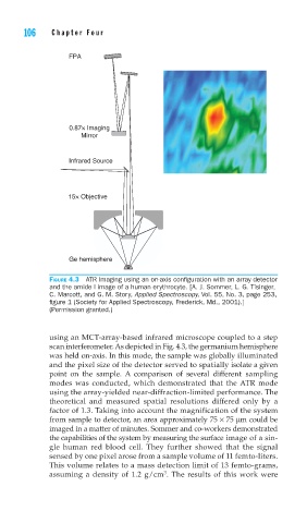
FIGURE 4.3 ATR Imaging using an on-axis confi guration with an array detector
and the amide I image of a human erythrocyte. [A. J. Sommer, L. G. Tisinger,
C. Marcott, and G. M. Story, Applied Spectroscopy, Vol. 55, No. 3, page 253,
fi gure 1 (Society for Applied Spectroscopy, Frederick, Md., 2001).]
(Permission granted.)
using an MCT-array-based infrared microscope coupled to a step
scan interferometer. As depicted in Fig. 4.3, the germanium hemisphere
was held on-axis. In this mode, the sample was globally illuminated
and the pixel size of the detector served to spatially isolate a given
point on the sample. A comparison of several different sampling
modes was conducted, which demonstrated that the ATR mode
using the array-yielded near-diffraction-limited performance. The
theoretical and measured spatial resolutions differed only by a
factor of 1.3. Taking into account the magnification of the system
from sample to detector, an area approximately 75 × 75 μm could be
imaged in a matter of minutes. Sommer and co-workers demonstrated
the capabilities of the system by measuring the surface image of a sin-
gle human red blood cell. They further showed that the signal
sensed by one pixel arose from a sample volume of 11 femto-liters.
This volume relates to a mass detection limit of 13 femto-grams,
3
assuming a density of 1.2 g/cm . The results of this work were