
Page 135 - Vibrational Spectroscopic Imaging for Biomedical Applications
P. 135
Evanescent W ave Imaging 111
1.1
1
0.9
0.8
Normalized Intensity 0.6
0.7
0.5
0.4
0.3
0.2
0.1
0
–0.1
–10 –8 –6 –4 –2 0 2 4 6 8 10
Micrometers (μm)
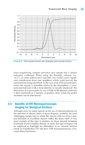
FIGURE 4.5 Point spread function and integrated point spread function.
from neighboring samples provided each sample had a similar
extinction coefficient. When using the Rayleigh criterion (i.e.,
“d/2”), as some authors have reported, one would expect signifi-
cant contributions from near neighbors which could prevent the
material from being identified. In the case of an ATR measurement,
where the sample is immobile relative to the hemisphere, a cross-
sectioned laminate with a sharp interface is usually employed. The
absorption of a given peak for one or both of the laminate materials
is then monitored as a function of position, from which the spatial
resolution can be determined.
4.5 Benefits of ATR Microspectroscopic
Imaging for Biological Sections
Although there are many reports on the use of infrared analysis for
the detection of disease states in tissue biopsies, probably the most
challenging sample type is where the disease state involves a min-
eral inclusion or crystalline deposit within the tissue itself. A very
good example of this type of situation are those mineral inclusions
commonly found in kidney disease. As such, this type of biopsy will
be employed to highlight the difficulties of an infrared analysis
based on transflection (TF) and how ATR microspectroscopy over-
comes those limitations.