
Page 131 - Vibrational Spectroscopic Imaging for Biomedical Applications
P. 131
Evanescent W ave Imaging 107
presented at the 2000 Pittsburgh Conference and later published
in Applied Spectroscopy. 30,31 In October of 2000, Biorad was issued
a patent for ATR imaging which was principally based on micro-
scopic on-axis measurements done with an array detector. 32
Although ATR imaging had been demonstrated, it was not
considered routine mainly due to the cost and complexity of the
associated step-scan interferometer and array detector. The neces-
sity to use a step-scan interferometer was a result of the relatively
33
slow read out capabilities of the MCT arrays. At FACSS in 2001,
Perkin Elmer introduced the Spotlight 300 infrared imaging
microscope which employed a linear array detector and a con-
ventional rapid scan interferometer. Perkin Elmer engineers
asked the question: “At what point does the size of the array dic-
tate the use of a step-scan interferometer?” They settled on a 16
element linear array. The so-called “push broom” mapping was
implemented through the careful synchronization of the detector,
“rapid” scan interferometer and the mapping stage. With this sys-
tem, off-axis ATR imaging could be conducted as proposed by Lewis
and Sommer. The next significant development came in 2006 when
Patterson and Havrilla realized that the spherical aberrations, which
limited the total sample area, were directly related to the radius of the
34
hemisphere. This realization was also made independently by Perkin
Elmer. Whereas Nakano and Kawata employed a 4-mm radius hemi-
sphere, Lewis and Sommer employed a 1.5-mm radius hemisphere,
Patterson and Havrilla employed a 12.5-mm radius (25-mm diameter)
germanium hemisphere. In conjunction with the off-axis scanning
on the Spotlight 300, the pair was able to obtain ATR images over an
area of 2500 × 2500 μm. The larger radius hemisphere also provided a
more constant penetration depth across the image, while maintaining
the spatial resolution. Patterson et al. later employed the same
hemisphere on a two-dimensional array system with a mapping
35
stage in the off-axis imaging mode. The basis for the experiment
was that the 4096 element array could generate images faster than a
16 element array. Their efforts produced marginal results due to the
fact that the image acquisition and stage synchronization was not
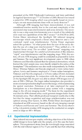
optimal among other factors. In 2006, Perkin Elmer developed and
introduced an ATR accessory based on the off-axis imaging concept
of Nakano and Kawata and Lewis and Sommer. The device shown
in Fig. 4.4 permits routine ATR imaging to be conducted on sample
areas as large as 400 × 400 μm.
4.4 Experimental Implementation
Most infrared microscopes employ reflecting objectives of the Schwar-
zchild design to focus light onto the sample, or in this case the hemi-
sphere. This requirement stems from the wavelength range associated
with the mid-infrared region (2.5 to 17 μm) and the fact that reflecting