
Page 170 - Vibrational Spectroscopic Imaging for Biomedical Applications
P. 170
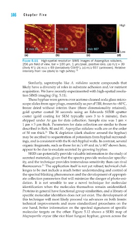
146    Cha pte r  F i v e


        FIGURE 5.11  High-spatial resolution SIMS images of Aspergillus nidulans,
        256 μm fi eld of view, bar = 100 μm, 1 μm/pixel, positive ions. (a) m/z = 39
                                       +
              +
        (likely K ); (b) m/z = 69 (tentatively C5H9 ), (c) m/z 607 (unknown). Relative
        intensity from low (dark) to high (white). 79

            Similarly, saprotrophs like  A. nidulans  secrete compounds that
        likely have a diversity of roles in substrate adhesion and/or nutrient
        acquisition. We have recently experimented with high-spatial resolu-
        tion SIMS imaging (Fig. 5.11).
            These hyphae were grown over acetone-cleaned soda glass micro-
        scope slides from agar plugs, essentially as per sFTIR; frozen to −80ºC,
        freeze dried without interim thaw (three dimensionality retained),
        gold sputter coated 30 seconds using an Edwards S150B sputter
        coater (gold coating for SEM typically uses 3 to 6 minute), then
        shipped under Ar gas for data collection. Sample size was 1 μm ×
        1 μm × 5 μm thick. Parameters for data collection are similar to those
        described in Refs. 80 and 81. Aspergillus nidulans walls are on the order
                     11
        of 50 nm thick.  The K depletion (dark shadow around the hyphae)
        may be ascribed to sequestration of potassium from hyphal surround-
        ings, and is consistent with the K-rich hyphal walls. In contrast, several
        organic fragments, such as those for m/z 69 and m/z 607 shown here,
        appear to be due to exudate secreted by growing hyphae.
            SERS can potentially provide valuable information in the study of
        secreted materials, given that the spectra provide molecular specific-
        ity, and the technique provides tremendous sensitivity than can rival
                   51
        fluorescence.  The application itself is not yet robust; technical chal-
        lenges to be met include a much better understanding and control of
        the spectral blinking phenomenon and the development of appropri-
        ate collection parameters that will minimize the danger of photo-oxi-
        dation. It is not sensible to use a novel technique for molecular
        identification when the molecules themselves remain unidentified.
        Proteins in general have functional group similarities, and a library of
        specific molecular identifiers does not yet exist. The development of
        this technique will most likely proceed via advances on both fronts:
        technical improvements and more standardized procedures on the
        one hand, better information on the spectral signatures of specific
        molecular targets on the other. Figure 5.12 shows a SERS map of
        Magnaporthe oryzae (the rice blast fungus) hyphae, grown across the