Page 261 - Vibrational Spectroscopic Imaging for Biomedical Applications
P. 261
Raman Micr oscopy for Biomedical Applications   237


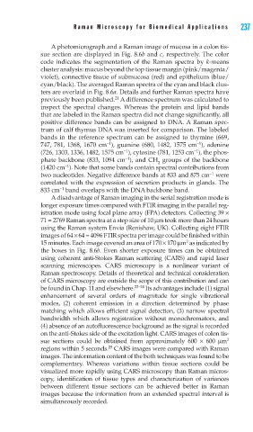
            A photomicrograph and a Raman image of mucosa in a colon tis-
        sue section are displayed in Fig. 8.6b and c, respectively. The color
        code indicates the segmentation of the Raman spectra by k-means
        cluster analysis: mucus beyond the top tissue margin (pink/magenta/
        violet), connective tissue of submucosa (red) and epithelium (blue/
        cyan/black). The averaged Raman spectra of the cyan and black clus-
        ters are overlaid in Fig. 8.6a. Details and further Raman spectra have
                               21
        previously been published.  A difference spectrum was calculated to
        inspect the spectral changes. Whereas the protein and lipid bands
        that are labeled in the Raman spectra did not change significantly, all
        positive difference bands can be assigned to DNA. A Raman spec-
        trum of calf thymus DNA was inserted for comparison. The labeled
        bands in the reference spectrum can be assigned to thymine (669,
                                                        −1
                             −1
        747, 781, 1368, 1670 cm ), guanine (680, 1482, 1575 cm ), adenine
                                  −1
                                                       −1
        (726, 1303, 1336, 1482, 1575 cm ), cytosine (781, 1253 cm ), the phos-
                                  −1
        phate backbone (833, 1094 cm ), and CH  groups of the backbone
                                            2
                −1
        (1420 cm ). Note that some bands contain spectral contributions from
                                                            −1
        two nucleotides. Negative difference bands at 833 and 875 cm  were
        correlated with the expression of secretion products in glands. The
              −1
        833 cm  band overlaps with the DNA backbone band.
            A disadvantage of Raman imaging in the serial registration mode is
        longer exposure times compared with FTIR imaging in the parallel reg-
        istration mode using focal plane array (FPA) detectors. Collecting 39 ×
        71 = 2769 Raman spectra at a step size of 10 μm took more than 24 hours
        using the Raman system Envia (Renishaw, UK). Collecting eight FTIR
        images of 64 × 64 = 4096 FTIR spectra per image could be finished within
                                                     2
        15 minutes. Each image covered an area of 170 × 170 μm  as indicated by
        the boxes in Fig. 8.6b. Even shorter exposure times can be obtained
        using coherent anti-Stokes Raman scattering (CARS) and rapid laser
        scanning microscopes. CARS microscopy is a nonlinear variant of
        Raman spectroscopy. Details of theoretical and technical consideration
        of CARS microscopy are outside the scope of this contribution and can
        be found in Chap. 11 and elsewhere. 22–24  Its advantages include (1) signal
        enhancement of several orders of magnitude for single vibrational
        modes, (2) coherent emission in a direction determined by phase
        matching which allows efficient signal detection, (3) narrow spectral
        bandwidth which allows registration without monochromators, and
        (4) absence of an autofluorescence background as the signal is recorded
        on the anti-Stokes side of the excitation light. CARS images of colon tis-
        sue sections could be obtained from approximately 600  × 600  μm 2
                             25
        regions within 5 seconds.  CARS images were compared with Raman
        images. The information content of the both techniques was found to be
        complementary. Whereas variations within tissue sections could be
        visualized more rapidly using CARS microscopy than Raman micros-
        copy, identification of tissue types and characterization of variances
        between different tissue sections can be achieved better in Raman
        images because the information from an extended spectral interval is
        simultaneously recorded.

