
Page 32 - Vibrational Spectroscopic Imaging for Biomedical Applications
P. 32
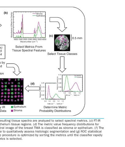
0.5 mm             Stroma  Epithelium  0.7  0.6  0.5  1556 cm –1   :  1652 cm –1
                              Select Tissue Classes  0.12  0.03  0.06  0.09  Relative Frequency  0.00  0.4  1.6  1.2


                    (c)                               0.8  0.4  0.0  1080 cm –1   :  1238 cm –1  Determine Metric Probability Distributions

         Stroma  Epithelium  4000 3000 3500  0.24  (d)  0.18  0.12  0.06  Relative Frequency  0.00

                      1000 1500 2000 2500 Wavenumber (cm –1 )  Select Metrics From Tissue Spectral Features  Epithelium  Stroma


        (b)          Absorbance (Offset for Clarity)             (a) FT-IR breast TMA image data is acquired and (b) the resulting tissue spectra are analyzed to select spectral metrics. (c) FT-IR  stroma and epithelium are determined and (e) each pixel on the spectral image of the breast TMA is classified a
                           ΔAUC  .645  .564  …  Sort Metrics by  ΔAUC and  Repeat  Classification  Classify IR  Image Data and H&E-stained images are then compared to select stroma and epithelium tissue regions. (d) The metric value frequency distributions for


                           Metric  1  2  …  (e)
                       (h)


                       Acquire IR images  1.00  0.99  0.98  0.97  Stroma  0.96  Epithelium  0.95  Mean  10  8  6  4  2  80  60  40  20  Number of Metrics Perform Statistical Analysis  (f)  Compare with  Gold Standard    converges at AUC ~ 1 at which (i) an optimal set of classification metrics is selected.


                               (g)  1.00  0.99  0.98  AUC  0.97  0.96  0.95
         0.3         0.0                       Obtain Final  Classification
        (a)
                                        (i)


                                                                 FIGURE 1.2


                                                                      9