
Page 77 - Vibrational Spectroscopic Imaging for Biomedical Applications
P. 77
Algal Cells, Cartilage, and IRENI 53
Monolayer
Absorbance
Standard Spectrum:
50% Proteoglycans, 30% M-CPPD,
20% BCP
3500 3000 2500 2000 1500 1000
–1
Wavenumbers (cm )
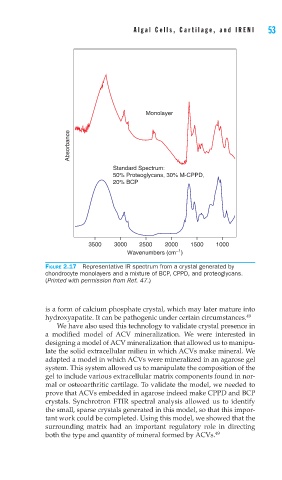
FIGURE 2.17 Representative IR spectrum from a crystal generated by
chondrocyte monolayers and a mixture of BCP, CPPD, and proteoglycans.
(Printed with permission from Ref. 47.)
is a form of calcium phosphate crystal, which may later mature into
hydroxyapatite. It can be pathogenic under certain circumstances. 49
We have also used this technology to validate crystal presence in
a modified model of ACV mineralization. We were interested in
designing a model of ACV mineralization that allowed us to manipu-
late the solid extracellular milieu in which ACVs make mineral. We
adapted a model in which ACVs were mineralized in an agarose gel
system. This system allowed us to manipulate the composition of the
gel to include various extracellular matrix components found in nor-
mal or osteoarthritic cartilage. To validate the model, we needed to
prove that ACVs embedded in agarose indeed make CPPD and BCP
crystals. Synchrotron FTIR spectral analysis allowed us to identify
the small, sparse crystals generated in this model, so that this impor-
tant work could be completed. Using this model, we showed that the
surrounding matrix had an important regulatory role in directing
both the type and quantity of mineral formed by ACVs. 49