
Page 75 - Vibrational Spectroscopic Imaging for Biomedical Applications
P. 75
Algal Cells, Cartilage, and IRENI 51
Synovial Fluid
Absorbance BCP Standard
Synovial Fluid
M-CPPD Standard
4000 3500 3000 2500 2000 1500 1000 500
–1
Wavenumbers (cm )
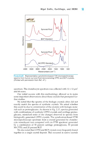
FIGURE 2.15 Representative synchrotron-based FTIR microspectroscopy
spectra from human synovial fl uid demonstrating CPPD and BCP crystals.
(Printed with permission from Ref. 47.)
spectrum. The chondrocyte spectrum was collected with 12 × 12 μm 2
and 64 scans.
Our initial success with this methodology allowed us to make
some important observations about these crystals that prompted fur-
ther studies.
We noted that the spectra of the biologic crystals often did not
exactly match the spectra of synthetic crystals. We asked whether
this could be due to contamination of the crystals with biologic mate-
rial such as proteoglycans. As shown in Fig. 2.17, spectra generated
with combinations of synthetic CPPD crystals and cartilage proteo-
glycans, mimicked some of the changes observed in spectra from
biologically generated CPPD crystals. The synchrotron-based FTIR
microspectroscopy spectrum from a crystal generated by chondro-
cyte monolayers was compared with an FTIR spectrum generated
by a combination of 50 percent cartilage proteoglycans, 30 percent
M-CPPD, and 20 percent BCP.
We also noted that CPPD and BCP crystals were frequently found
together in a single crystal deposit. This occurred in native crystals