
Page 78 - Vibrational Spectroscopic Imaging for Biomedical Applications
P. 78
54 Cha pte r T w o
Absorbance M-CPPD Absorbance M-CPPD Absorbance Point 3
Point 2
M-CPPD
Point 1
4000 3000 2000 1000 4000 3000 2000 1000 4000 3000 2000 1000
–1
–1
–1
Wavenumbers (cm ) Wavenumbers (cm ) Wavenumbers (cm )
Panel A Panel B Panel C
6 4 5 3 2 1
Integrated over 985–1155 cm –1 Integrated over 1125–1201 cm –1
Absorbance Point 4 Absorbance Point 5 Absorbance Point 6
BCP BCP BCP
4000 3000 2000 1000 4000 3000 2000 1000 4000 3000 2000 1000
–1
–1
–1
Wavenumbers (cm ) Wavenumbers (cm ) Wavenumbers (cm )
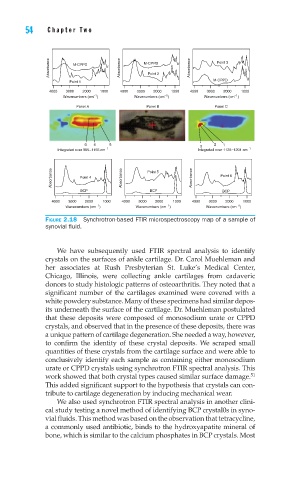
FIGURE 2.18 Synchrotron-based FTIR microspectroscopy map of a sample of
synovial fl uid.
We have subsequently used FTIR spectral analysis to identify
crystals on the surfaces of ankle cartilage. Dr. Carol Muehleman and
her associates at Rush Presbyterian St. Luke’s Medical Center,
Chicago, Illinois, were collecting ankle cartilages from cadaveric
donors to study histologic patterns of osteoarthritis. They noted that a
significant number of the cartilages examined were covered with a
white powdery substance. Many of these specimens had similar depos-
its underneath the surface of the cartilage. Dr. Muehleman postulated
that these deposits were composed of monosodium urate or CPPD
crystals, and observed that in the presence of these deposits, there was
a unique pattern of cartilage degeneration. She needed a way, however,
to confirm the identity of these crystal deposits. We scraped small
quantities of these crystals from the cartilage surface and were able to
conclusively identify each sample as containing either monosodium
urate or CPPD crystals using synchrotron FTIR spectral analysis. This
work showed that both crystal types caused similar surface damage. 51
This added significant support to the hypothesis that crystals can con-
tribute to cartilage degeneration by inducing mechanical wear.
We also used synchrotron FTIR spectral analysis in another clini-
cal study testing a novel method of identifying BCP crystal0s in syno-
vial fluids. This method was based on the observation that tetracycline,
a commonly used antibiotic, binds to the hydroxyapatite mineral of
bone, which is similar to the calcium phosphates in BCP crystals. Most