
Page 385 - Advances in Biomechanics and Tissue Regeneration
P. 385
382 19. IMPACT OF MECHANOBIOLOGICAL PERTURBATION IN CARTILAGE TISSUE ENGINEERING
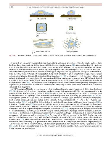
FIG. 19.3 Schematic diagrams of the response of cells to substrates with different stiffness (A), surface area (B), and topography (C).
Stem cells are exquisitely sensitive to the biochemical and mechanical properties of the extracellular matrix, which
has been shown to regulate the differentiation of MSCs towards specific lineages [45]. When cultured on 2-D substrates
that mimicked the stiffness of physiologic tissue environments, MSCs adopted a phenotype corresponding to the tissue
stiffness, as demonstrated by cellular morphology, transcript markers, and protein production. In 2-D culture systems,
substrate stiffness generally affects cellular morphology. Compared with myogenic and osteogenic differentiation,
MSC chondrogenesis preferred softer substratum that permits adoption of spherical cell morphology, with lower cell
adhesion strength and decreased F-actin stress fiber formation [46–48]. Investigation of both substrate stiffness and
adhesivity (through provision of Arg-Gly-Asp peptide, RGD) on electrospun hyaluronic acid (HA) fibers indicated
that MSC spreading and focal adhesion formation were dependent on RGD density, with traction force increased with
more adhesive fibers [49]. The expression of chondrogenic markers, unlike trends in cell spreading and cytoskeletal
organization, was influenced by both fiber mechanics and adhesivity in which softer fibers and lower RGD densities
enhanced chondrogenesis.
In 3-D hydrogels, MSCs have been shown to retain a spherical morphology irrespective of the hydrogel stiffness
[50, 51]. A study in 3-D hydrogel found that modulus-driven differentiation of MSCs was independent of actin
polymerization, ROCK signaling, or NMM II [51]. In spite of this, the fate of encapsulated MSCs is still dependent
on the stiffness of the hydrogel, with a decrease of chondrogenesis with increasing gel stiffness [42, 47, 52, 53].
Development of HA-based scaffolds with tunable mechanical and rheological properties has allowed the identi-
fication of an optimal, lower cross-linked matrix elasticity (Young’smodulus at 3–6kPa) to favor hyaline carti-
lage formation [53]. A shift in MSC differentiation towards the fibrocartilage and fibrous tissue formation [52] or
induction of calcification [53] was reported with increasing cross-linking and matrix stiffness of HA hydrogels.
Substrates with compliant mechanical cues can also facilitate matrix-induced cell-cell interactions that are essential
for the recapitulation of precartilage mesenchymal condensation [32, 54].Engagement withintegrinwas shownto
be essential for MSC chondrogenesisasbothcollagenand RGD-modified hydrogel enhanced differentiation [55–
57]. Prior to condensation, cell-matrix interactions in hyaluronan and type I collagen (Col I)–rich ECM mediate
aggregation of mesenchymal progenitors [58]. Provision of a 3-D environment with RGD, hyaluronic acid,
and/or type I collagen, synergized with a softer hydrogel to support MSC condensation, enhanced cartilaginous
development [57]. Mechanosensing computational models have been designed to relate the role of substratum
mechanical cues in directing MSC proliferation and differentiation in 3-D hydrogel and could be employed to pre-
dict essential aspects of cell maturation, differentiation, proliferation and apoptosis during regenerative events
[59–61]. Such computational models could be favorably exploited to identify substratum mechano-specificity in
tissue engineering applications.
II. MECHANOBIOLOGY AND TISSUE REGENERATION