
Page 102 - Artificial Intelligence for Computational Modeling of the Heart
P. 102
72 Chapter 2 Implementation of a patient-specific cardiac model
Figure 2.22. The effect of arteries and atria on the ventricles is modeled by
attaching springs to the valve plane.
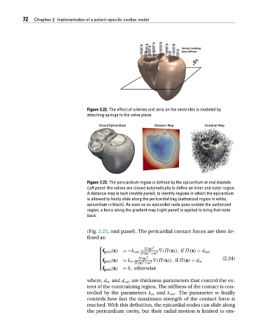
Figure 2.23. The pericardium region is defined by the epicardium at end diastole.
Left panel: the valves are closed automatically to define an inner and outer region.
A distance map is built (middle panel), to identify regions in which the epicardium
is allowed to freely slide along the pericardial bag (authorized region in white,
epicardium in black). As soon as an epicardial node goes outside the authorized
region, a force along the gradient map (right panel) is applied to bring that node
back.
(Fig. 2.23, mid panel). The pericardial contact forces are then de-
fined as:
⎧ 2
Π(x)
⎪f peri (x) =−k out Π(x) +m 2 ∇(Π(x)), if Π(x)>d out
⎪
2
⎨ Π(x) 2
f
⎪ peri (x) = k in Π(x) +m 2 ∇(Π(x)), if Π(x)<d in (2.24)
2
⎪
f peri (x) = 0, otherwise
⎩
where, d in and d out are thickness parameters that control the ex-
tent of the constraining region. The stiffness of the contact is con-
trolled by the parameters k in and k out .The parameter m finally
controls how fast the maximum strength of the contact force is
reached. With this definition, the epicardial nodes can slide along
the pericardium cavity, but their radial motion is limited to em-