
Page 77 - Artificial Intelligence for Computational Modeling of the Heart
P. 77
Chapter 2 Implementation of a patient-specific cardiac model 47
the meshing techniques would become more involved, in particu-
lar when the underlying shape is as complex as a patient-specific,
diseased heart. The anatomical structures are automatically anno-
tated on the tetrahedral mesh by leveraging the parameterization
of the segmented surfaces (Fig. 2.5). Myocardium scar or fibro-
sis, identified in images like delayed-enhancement MRI, are also
mapped onto the anatomical model to account for their impact
on cardiac function.
Figure 2.5. Tagged surface meshes (left and middle panels) and fused tetrahedral
mesh (right panel).
The fibrous, collageneous tissue is also tagged on the model,
with the aim to properly represent its inactive electro-mechanical
properties. Based on literature reports, a rule-based classification
of fibrous tissue can be designed. This comprises the fibrous rings
of the pulmonary and aortic valves, as well as fibrous connec-
tions linking these rings to the atrioventricular valves [204,205].
We therefore tag as fibrous tissue all mesh elements in the left
ventricular outflow tract as well as all mesh elements in the right
ventricular outflow tract and above the plane of the atrioventric-
ular valves (Fig. 2.5). Finally, for regional parameter estimation or
function analysis, a subdivision in segments (according to the def-
inition proposed in [206]) is automatically defined based on the
tags, as illustrated in Fig. 2.6.
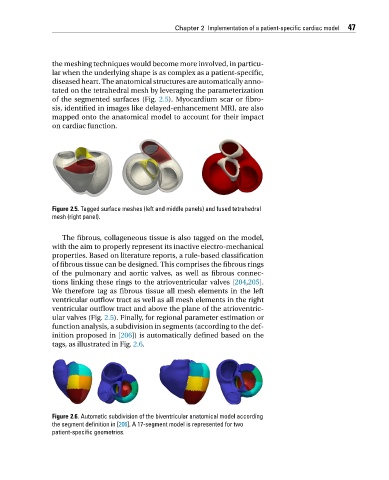
Figure 2.6. Automatic subdivision of the biventricular anatomical model according
the segment definition in [206]. A 17-segment model is represented for two
patient-specific geometries.