
Page 74 - Artificial Intelligence for Computational Modeling of the Heart
P. 74
44 Chapter 2 Implementation of a patient-specific cardiac model
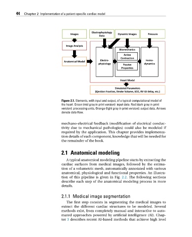
Figure 2.1. Elements, with input and output, of a typical computational model of
the heart. Green (mid gray in print version): input data. Red (dark gray in print
version): processing units. Orange (light gray in print version): output data. Arrows
denote data flow.
mechano-electrical feedback (modification of electrical conduc-
tivity due to mechanical pathologies) could also be modeled if
required by the application. This chapter provides implementa-
tion details of each component, knowledge that will be needed for
the remainder of the book.
2.1 Anatomical modeling
A typical anatomical modeling pipeline starts by extracting the
cardiac surfaces from medical images, followed by the estima-
tion of a volumetric mesh, automatically annotated with various
anatomical, physiological and functional properties. An illustra-
tion of this pipeline is given in Fig. 2.2. The following sections
describe each step of the anatomical modeling process in more
details.
2.1.1 Medical image segmentation
The first step consists in segmenting the medical images to
extract the different cardiac structures to be modeled. Several
methods exist, from completely manual and interactive to auto-
mated approaches powered by artificial intelligence (AI). Chap-
ter 3 describes recent AI-based methods that achieve high level