
Page 119 - Biomedical Engineering and Design Handbook Volume 1, Fundamentals
P. 119
96 BIOMECHANICS OF THE HUMAN BODY
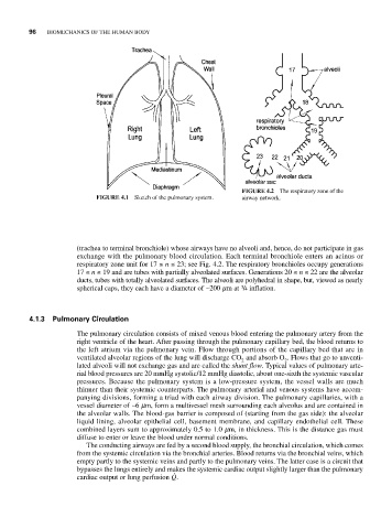
FIGURE 4.2 The respiratory zone of the
FIGURE 4.1 Sketch of the pulmonary system. airway network.
(trachea to terminal bronchiole) whose airways have no alveoli and, hence, do not participate in gas
exchange with the pulmonary blood circulation. Each terminal bronchiole enters an acinus or
respiratory zone unit for 17 ≤ n ≤ 23; see Fig. 4.2. The respiratory bronchioles occupy generations
17 ≤ n ≤ 19 and are tubes with partially alveolated surfaces. Generations 20 ≤ n ≤ 22 are the alveolar
ducts, tubes with totally alveolated surfaces. The alveoli are polyhedral in shape, but, viewed as nearly
spherical caps, they each have a diameter of ~200 mm at 3 /4 inflation.
4.1.3 Pulmonary Circulation
The pulmonary circulation consists of mixed venous blood entering the pulmonary artery from the
right ventricle of the heart. After passing through the pulmonary capillary bed, the blood returns to
the left atrium via the pulmonary vein. Flow through portions of the capillary bed that are in
ventilated alveolar regions of the lung will discharge CO and absorb O . Flows that go to unventi-
2
2
lated alveoli will not exchange gas and are called the shunt flow. Typical values of pulmonary arte-
rial blood pressures are 20 mmHg systolic/12 mmHg diastolic, about one-sixth the systemic vascular
pressures. Because the pulmonary system is a low-pressure system, the vessel walls are much
thinner than their systemic counterparts. The pulmonary arterial and venous systems have accom-
panying divisions, forming a triad with each airway division. The pulmonary capillaries, with a
vessel diameter of ~6 mm, form a multivessel mesh surrounding each alveolus and are contained in
the alveolar walls. The blood-gas barrier is composed of (starting from the gas side): the alveolar
liquid lining, alveolar epithelial cell, basement membrane, and capillary endothelial cell. These
combined layers sum to approximately 0.5 to 1.0 mm, in thickness. This is the distance gas must
diffuse to enter or leave the blood under normal conditions.
The conducting airways are fed by a second blood supply, the bronchial circulation, which comes
from the systemic circulation via the bronchial arteries. Blood returns via the bronchial veins, which
empty partly to the systemic veins and partly to the pulmonary veins. The latter case is a circuit that
bypasses the lungs entirely and makes the systemic cardiac output slightly larger than the pulmonary
cardiac output or lung perfusion Q.