
Page 120 - Biomedical Engineering and Design Handbook Volume 1, Fundamentals
P. 120
RESPIRATORY MECHANICS AND GAS EXCHANGE 97
4.1.4 Lymphatics and Nerves
To maintain homeostasis, the continuous transudation of fluid and solutes from the pulmonary cap-
illary bed into the surrounding interstitium and alveolar space is balanced by lymphatic drainage out
of the lung. The lymphatic flow is directed toward the hilum from the pleural surfaces. From lymph
nodes in the hilum, the lymph travels to the paratracheal nodes and then eventually into the venous
system via the thoracic duct. The lung has nerve fibers from both the vagal nerves (parasympathetic)
and the sympathetic nerves. The efferent fibers go to the bronchial musculature and the afferents
come from the bronchi and alveoli.
4.2 MECHANICS OF BREATHING
4.2.1 Chest Wall
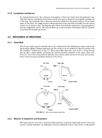
The rib cage and its muscles form the chest wall, which protects the vital thoracic organs while keep-
ing the lungs inflated. During inspiration, the ribs swing on an axis defined by their articulation with
the vertebrae, 20 dashed lines in Fig. 4.3. The result is that upper ribs, such as rib 1, swing forward
and up, like a pump handle, increasing the anterior-posterior diameter of the upper chest wall.
However, lower ribs swing primarily laterally, like a bucket handle pinned at the spine and sternum.
Their motion increases the lateral diameter of the thorax.
FIGURE 4.3 Rib motion during inspiration.
4.2.2 Muscles of Inspiration and Expiration
The major muscles involved in inspiration and expiration include the diaphragm and the intercostal
muscles. During inspiration, the diaphragm contracts, pulling the inferior lung surface (via the pleural