
Page 121 - Biomedical Engineering and Design Handbook Volume 1, Fundamentals
P. 121
98 BIOMECHANICS OF THE HUMAN BODY
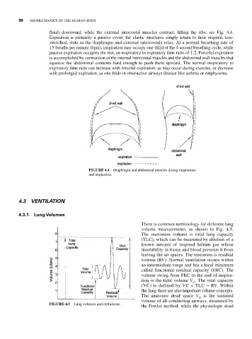
fluid) downward, while the external intercostal muscles contract, lifting the ribs; see Fig. 4.4.
Expiration is primarily a passive event; the elastic structures simply return to their original, less-
stretched, state as the diaphragm and external intercostals relax. At a normal breathing rate of
15 breaths per minute (bpm), inspiration may occupy one-third of the 4-second breathing cycle, while
passive expiration occupies the rest, an inspiratory to expiratory time ratio of 1:2. Forceful expiration
is accomplished by contraction of the internal intercostal muscles and the abdominal wall muscles that
squeeze the abdominal contents hard enough to push them upward. The normal inspiratory to
expiratory time ratio can increase with forceful expiration, as may occur during exercise, or decrease
with prolonged expiration, as one finds in obstructive airways disease like asthma or emphysema.
FIGURE 4.4 Diaphragm and abdominal muscles during inspiration
and expiration.
4.3 VENTILATION
4.3.1 Lung Volumes
There is common terminology for different lung
volume measurements, as shown in Fig. 4.5.
The maximum volume is total lung capacity
(TLC), which can be measured by dilution of a
known amount of inspired helium gas whose
insolubility in tissue and blood prevents it from
leaving the air spaces. The minimum is residual
volume (RV). Normal ventilation occurs within
an intermediate range and has a local minimum
called functional residual capacity (FRC). The
volume swing from FRC to the end of inspira-
tion is the tidal volume V . The vital capacity
T
(VC) is defined by VC = TLC − RV. Within
the lung there are also important volume concepts.
The anatomic dead space V D is the summed
volume of all conducting airways, measured by
FIGURE 4.5 Lung volumes and definitions.
the Fowler method, while the physiologic dead