
Page 205 - Biomedical Engineering and Design Handbook Volume 1, Fundamentals
P. 205
182 BIOMECHANICS OF THE HUMAN BODY
400 10000
pAC
8000
Ligament force (N) 200 aAC aPC 6000 Muscle force (N)
300
Quads
4000
100
pPC 2000
0 0
0 30 60 90
Knee flexion (deg)
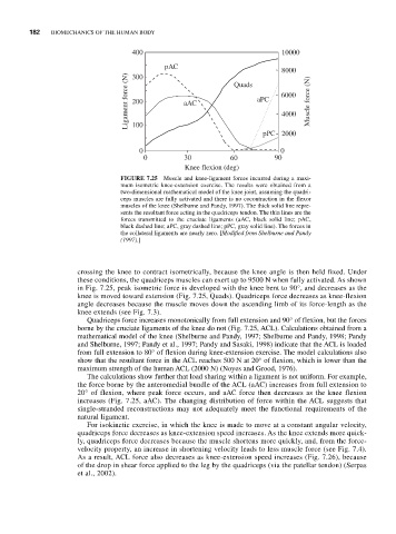
FIGURE 7.25 Muscle and knee-ligament forces incurred during a maxi-
mum isometric knee-extension exercise. The results were obtained from a
two-dimensional mathematical model of the knee joint, assuming the quadri-
ceps muscles are fully activated and there is no cocontraction in the flexor
muscles of the knee (Shelburne and Pandy, 1997). The thick solid line repre-
sents the resultant force acting in the quadriceps tendon. The thin lines are the
forces transmitted to the cruciate ligaments (aAC, black solid line; pAC,
black dashed line; aPC, gray dashed line; pPC, gray solid line). The forces in
the collateral ligaments are nearly zero. [Modified from Shelburne and Pandy
(1997).]
crossing the knee to contract isometrically, because the knee angle is then held fixed. Under
these conditions, the quadriceps muscles can exert up to 9500 N when fully activated. As shown
in Fig. 7.25, peak isometric force is developed with the knee bent to 90°, and decreases as the
knee is moved toward extension (Fig. 7.25, Quads). Quadriceps force decreases as knee-flexion
angle decreases because the muscle moves down the ascending limb of its force-length as the
knee extends (see Fig. 7.3).
Quadriceps force increases monotonically from full extension and 90° of flexion, but the forces
borne by the cruciate ligaments of the knee do not (Fig. 7.25, ACL). Calculations obtained from a
mathematical model of the knee (Shelburne and Pandy, 1997; Shelburne and Pandy, 1998; Pandy
and Shelburne, 1997; Pandy et al., 1997; Pandy and Sasaki, 1998) indicate that the ACL is loaded
from full extension to 80° of flexion during knee-extension exercise. The model calculations also
show that the resultant force in the ACL reaches 500 N at 20° of flexion, which is lower than the
maximum strength of the human ACL (2000 N) (Noyes and Grood, 1976).
The calculations show further that load sharing within a ligament is not uniform. For example,
the force borne by the anteromedial bundle of the ACL (aAC) increases from full extension to
20° of flexion, where peak force occurs, and aAC force then decreases as the knee flexion
increases (Fig. 7.25, aAC). The changing distribution of force within the ACL suggests that
single-stranded reconstructions may not adequately meet the functional requirements of the
natural ligament.
For isokinetic exercise, in which the knee is made to move at a constant angular velocity,
quadriceps force decreases as knee-extension speed increases. As the knee extends more quick-
ly, quadriceps force decreases because the muscle shortens more quickly, and, from the force-
velocity property, an increase in shortening velocity leads to less muscle force (see Fig. 7.4).
As a result, ACL force also decreases as knee-extension speed increases (Fig. 7.26), because
of the drop in shear force applied to the leg by the quadriceps (via the patellar tendon) (Serpas
et al., 2002).