
Page 209 - Biomedical Engineering and Design Handbook Volume 1, Fundamentals
P. 209
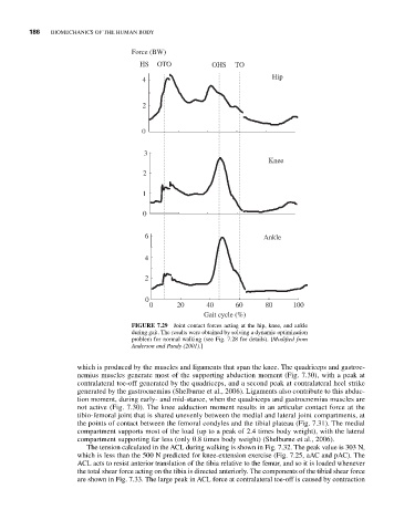
186  BIOMECHANICS OF THE HUMAN BODY

                                    Force (BW)
                                      HS   OTO          OHS   TO
                                                                       Hip
                                       4

                                       2


                                       0


                                       3
                                                                      Knee
                                       2

                                       1

                                       0

                                        6                            Ankle


                                        4

                                        2

                                        0
                                         0      20     40     60      80     100
                                                      Gait cycle (%)
                                    FIGURE 7.29  Joint contact forces acting at the hip, knee, and ankle
                                    during gait. The results were obtained by solving a dynamic optimization
                                    problem for normal walking (see Fig. 7.28 for details). [Modified from
                                    Anderson and Pandy (2001).]

                       which is produced by the muscles and ligaments that span the knee. The quadriceps and gastroc-
                       nemius muscles generate most of the supporting abduction moment (Fig. 7.30), with a peak at
                       contralateral toe-off generated by the quadriceps, and a second peak at contralateral heel strike
                       generated by the gastrocnemius (Shelburne et al., 2006). Ligaments also contribute to this abduc-
                       tion moment, during early- and mid-stance, when the quadriceps and gastrocnemius muscles are
                       not active (Fig. 7.30). The knee adduction moment results in an articular contact force at the
                       tibio-femoral joint that is shared unevenly between the medial and lateral joint compartments, at
                       the points of contact between the femoral condyles and the tibial plateau (Fig. 7.31). The medial
                       compartment supports most of the load (up to a peak of 2.4 times body weight), with the lateral
                       compartment supporting far less (only 0.8 times body weight) (Shelburne et al., 2006).
                         The tension calculated in the ACL during walking is shown in Fig. 7.32. The peak value is 303 N,
                       which is less than the 500 N predicted for knee-extension exercise (Fig. 7.25, aAC and pAC). The
                       ACL acts to resist anterior translation of the tibia relative to the femur, and so it is loaded whenever
                       the total shear force acting on the tibia is directed anteriorly. The components of the tibial shear force
                       are shown in Fig. 7.33. The large peak in ACL force at contralateral toe-off is caused by contraction