
Page 211 - Biomedical Engineering and Design Handbook Volume 1, Fundamentals
P. 211
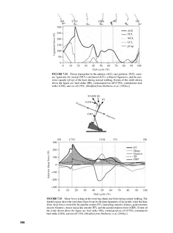
HS    CTO            CHS      TO           HS
                                    300
                                                                            ACL
                                    250
                                   Ligament force (N)  200                  MCL
                                                                            PCL
                                                                            LCL
                                    150
                                                                            pCap
                                    100
                                    50
                                     0
                                       0   10  20  30   40  50  60   70  80  90  100
                                                        Gait cycle (%)
                                 FIGURE 7.32  Forces transmitted to the anterior (ACL) and posterior (PCL) cruci-
                                 ate ligaments, the medial (MCL) and lateral (LCL) collateral ligaments, and the pos-
                                 terior capsule (pCap) of the knee during normal walking. Events of the stride shown
                                 above the figure are: heel strike (HS), contralateral toe-off (CTO), contralateral heel
                                 strike (CHS), and toe-off (TO). [Modified from Shelburne et al. (2004a).]


                                                     HAMS TF
                                                             PT
                                                    GAS
                                              Posterior
                                                     CRF        Anterior


                                    HS     CTO              CHS     TO               HS
                                 300
                                                                               PT
                                 200                                           Hams
                               Anterior shear force (N)  –100                  GRF
                                                                               Gastroc
                                                                               TF
                                 100
                                   0


                                –200
                                –300
                                    0    10   20   30   40   50   60  70   80   90   100
                                                         Gait cycle (%)
                              FIGURE 7.33 Shear forces acting on the lower leg (shank and foot) during normal walking. The
                              shaded region shows the total shear force borne by the knee ligaments of the model, while the lines
                              show shear forces exerted by the patellar tendon (PT), hamstrings muscles (Hams), gastrocnemius
                              muscle (Gastroc), tensor fascia lata muscle (TF), and the ground reaction force (GRF). Events of
                              the stride shown above the figure are: heel strike (HS), contralateral toe-off (CTO), contralateral
                              heel strike (CHS), and toe-off (TO). [Modified from Shelburne et al. (2004a).]

               188