
Page 258 - Biomedical Engineering and Design Handbook Volume 1, Fundamentals
P. 258
BONE MECHANICS 235
Criteria such as the Tsai-Wu criterion have only a lim-
ited ability to describe multiaxial failure of trabecular
bone for arbitrary stress states. Coupling between nor-
mal strengths in different directions (longitudinal ver-
sus transverse, for example) appears to be minimal. 104
At present, it is recommended for multiaxial failure
analysis that the tensile-compressive-shear-strength
asymmetry be recognized, as well as the strength
anisotropy. If properties are not available for a specific
site under analysis, failure strains should be used from
sites that have a similar density range and architecture.
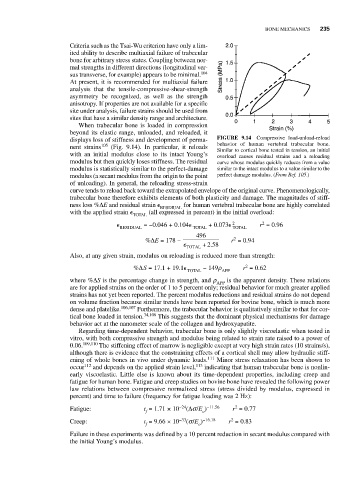
When trabecular bone is loaded in compression
beyond its elastic range, unloaded, and reloaded, it
displays loss of stiffness and development of perma- FIGURE 9.14 Compressive load-unload-reload
behavior of human vertebral trabecular bone.
nent strains 105 (Fig. 9.14). In particular, it reloads Similar to cortical bone tested in tension, an initial
with an initial modulus close to its intact Young’s overload causes residual strains and a reloading
modulus but then quickly loses stiffness. The residual curve whose modulus quickly reduces from a value
modulus is statistically similar to the perfect-damage similar to the intact modulus to a value similar to the
modulus (a secant modulus from the origin to the point perfect damage modulus. (From Ref. 105.)
of unloading). In general, the reloading stress-strain
curve tends to reload back toward the extrapolated envelope of the original curve. Phenomenologically,
trabecular bone therefore exhibits elements of both plasticity and damage. The magnitudes of stiff-
ness loss %ΔE and residual strain RESDIUAL for human vertebral trabecular bone are highly correlated
with the applied strain (all expressed in percent) in the initial overload:
TOTAL
2
=−0.046 + 0.104 + 0.073 2 r = 0.96
RESIDUAL TOTAL TOTAL
496
%ΔE = 178 − r = 0.94
2
+ 258
.
TOTAL
Also, at any given strain, modulus on reloading is reduced more than strength:
2
%ΔS = 17.1 + 19.1 − 149r r = 0.62
TOTAL APP
where %ΔS is the percentage change in strength, and r APP is the apparent density. These relations
are for applied strains on the order of 1 to 5 percent only; residual behavior for much greater applied
strains has not yet been reported. The percent modulus reductions and residual strains do not depend
on volume fraction because similar trends have been reported for bovine bone, which is much more
dense and platelike. 106,107 Furthermore, the trabecular behavior is qualitatively similar to that for cor-
tical bone loaded in tension. 34,108 This suggests that the dominant physical mechanisms for damage
behavior act at the nanometer scale of the collagen and hydroxyapatite.
Regarding time-dependent behavior, trabecular bone is only slightly viscoelastic when tested in
vitro, with both compressive strength and modulus being related to strain rate raised to a power of
0.06. 109,110 The stiffening effect of marrow is negligible except at very high strain rates (10 strains/s),
although there is evidence that the constraining effects of a cortical shell may allow hydraulic stiff-
ening of whole bones in vivo under dynamic loads. 111 Minor stress relaxation has been shown to
occur 112 and depends on the applied strain level, 113 indicating that human trabecular bone is nonlin-
early viscoelastic. Little else is known about its time-dependent properties, including creep and
fatigue for human bone. Fatigue and creep studies on bovine bone have revealed the following power
law relations between compressive normalized stress (stress divided by modulus, expressed in
percent) and time to failure (frequency for fatigue loading was 2 Hz):
2
Fatigue: t = 1.71 × 10 −24 (Δs/E ) −11.56 r = 0.77
f o
2
Creep: t = 9.66 × 10 −33 (s/E ) −16.18 r = 0.83
f o
Failure in these experiments was defined by a 10 percent reduction in secant modulus compared with
the initial Young’s modulus.