Page 295 - Color Atlas of Biochemistry
P. 295
286       Tissues and organs


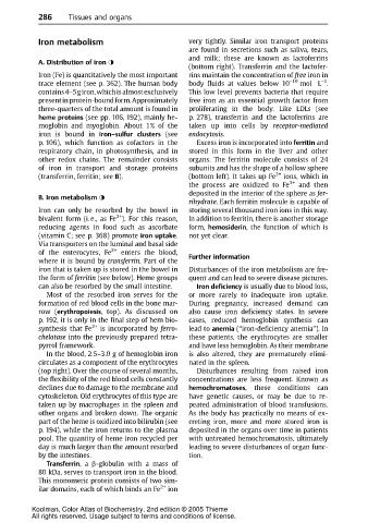
             Iron metabolism                                  very tightly. Similar iron transport proteins
                                                              are found in secretions such as saliva, tears,
                                                              and milk; these are known as lactoferrins
             A. Distribution of iron
                                                              (bottom right). Transferrin and the lactofer-
             Iron (Fe) is quantitatively the most important   rins maintain the concentration of free iron in
                                                                                                         –1
             trace element (see p. 362). The human body       body fluidsatvaluesbelow 10     –10  mol  L .
             contains4–5giron,whichisalmostexclusively        This low level prevents bacteria that require
             presentinprotein-boundform.Approximately         free iron as an essential growth factor from
             three-quarters of the total amount is found in   proliferating in the body. Like LDLs (see
             heme proteins (see pp. 106, 192), mainly he-     p. 278), transferrin and the lactoferrins are
             moglobin and myoglobin. About 1% of the          taken up into cells by receptor-mediated
             iron is bound in iron–sulfur clusters (see       endocytosis.
             p. 106), which function as cofactors in the         Excess iron is incorporated into ferritin and
             respiratory chain, in photosynthesis, and in     stored in this form in the liver and other
             other redox chains. The remainder consists       organs. The ferritin molecule consists of 24
             of iron in transport and storage proteins        subunits and has the shape of a hollow sphere
             (transferrin, ferritin; see B).                  (bottom left). It takes up Fe 2+  ions, which in
                                                              the process are oxidized to Fe  3+  and then
                                                              deposited in the interior of the sphere as fer-
             B. Iron metabolism
                                                              rihydrate. Each ferritin molecule is capable of
             Iron can onlybe resorbedbythe bowel in           storing several thousand iron ions in this way.
                                       2+
             bivalent form (i. e., as Fe ). For this reason,  In addition to ferritin, there is another storage
             reducing agents in food such as ascorbate        form, hemosiderin, the function of which is
             (vitamin C; see p. 368) promote iron uptake.     not yet clear.
             Via transporters on the luminal and basal side
             of the enterocytes, Fe  2+  enters the blood,
             where it is bound by transferrin. Part of the    Further information
             iron that is taken up is stored in the bowel in  Disturbances of the iron metabolism are fre-
             the form of ferritin (see below). Heme groups    quent and can lead to severe disease pictures.
             can also be resorbed by the small intestine.        Iron deficiency is usually due to blood loss,
                Most of the resorbed iron serves for the      or more rarely to inadequate iron uptake.
             formation of red blood cells in the bone mar-    During pregnancy, increased demand can
             row (erythropoiesis,top). As discussed on        also cause iron deficiency states. In severe
             p. 192, it is only in the final step of hem bio-  cases, reduced hemoglobin synthesis can
             synthesis that Fe 2+  is incorporated by ferro-  lead to anemia (“iron-deficiency anemia”). In
             chelatase into the previously prepared tetra-    thesepatients, theerythrocytes are smaller
             pyrrol framework.                                and have less hemoglobin. As their membrane
                In the blood, 2.5–3.0 g of hemoglobin iron    is also altered, they are prematurely elimi-
             circulates as a component of the erythrocytes    nated in the spleen.
             (top right). Over the course of several months,     Disturbances resulting from raised iron
             the flexibility of the red blood cells constantly  concentrations are less frequent. Known as
             declines due to damage to the membrane and       hemochromatoses,     these   conditions  can
             cytoskeleton. Old erythrocytes of this type are  have genetic causes, or may be due to re-
             taken up by macrophages in the spleen and        peated administration of blood transfusions.
             other organs and broken down. The organic        As the body has practically no means of ex-
             part of the heme is oxidized into bilirubin (see  creting iron, more and more stored iron is
             p. 194), while the iron returns to the plasma    deposited in the organs over time in patients
             pool. The quantity of heme iron recycled per     with untreated hemochromatosis, ultimately
             day is much larger than the amount resorbed      leading to severe disturbances of organ func-
             by the intestines.                               tion.
                Transferrin,a β-globulin with a mass of
             80 kDa, serves to transport iron in the blood.
             This monomeric protein consists of two sim-
             ilar domains, each of which binds an Fe 2+  ion


           Koolman, Color Atlas of Biochemistry, 2nd edition © 2005 Thieme
           All rights reserved. Usage subject to terms and conditions of license.

