
Page 172 - Computational Modeling in Biomedical Engineering and Medical Physics
P. 172
Bioimpedance methods 161
without loss of accuracy, the AC model may be replaced with a simpler DC model. This
may be explained by the irrotationality of the electric field and the apparent disparity
between the operating frequency of the ECM and the hemodynamic of the time scale.
A key problem of the electromechanic coupling is the blood electrical conductivity.
This is solved here with an equivalent quantity, calculated out of analytical expressions
using averaging methods. However, some electrophysiology effects are not evidenced
when embracing the numerical model, for example, the nonlinear change in the blood
electrical conductivity that happens for increasing and decreasing the flow rates. Even so,
the sensitivity of the solution to blood flow pulsations is evidently outlined.
The ECM impedance and its derivative with respect to time found by numerical
modelization fit fairly well with the experimental results. Significant cardio-hemodynamic
indices, consistent with experimental findings, are evidenced by the numerical results.
Several cardiovascular indices of importance in medical diagnosis become thus mathemati-
cally tractable, for example: the start of blood ejection by the left ventricle, the systolic
major upward deflection, the aortic valve closure, the diastolic upward deflection, the left-
ventricular ejection time, and dZ/dt max .
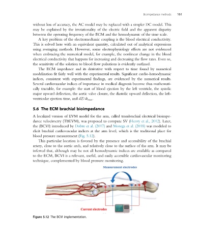
5.6 The ECM brachial bioimpedance
A localized version of EVM model for the arm, called transbrachial electrical bioimpe-
dance velocimetry (TBEVM), was proposed to compute SV (Henry et al., 2012). Later,
the (BCVI) introduced by Dobre et al. (2017) and Morega et al. (2018) was modeled to
elicit brachial cardiovascular indices at the arm level, which is the traditional place for
blood pressure measurement (Fig. 5.12).
This particular location is favored by the presence and accessibility of the brachial
artery, close to the aortic arch, and relatively close to the surface of the arm. It may be
inferred that, although may be not all hemodynamic indices are available as compared
to the ECM, BCVI is a relevant, useful, and easily accessible cardiovascular monitoring
technique, complemented by blood pressure monitoring.
Figure 5.12 The BCVI implementation.