Page 129 - Computational Retinal Image Analysis
P. 129

122    CHAPTER 7  OCT layer segmentation


                          Table 1  Description of major retinal layers and their functioning.
                          Layer name                        Function
                          Nerve fiber layer (NFL)           Axons of ganglion cells
                          Ganglion cell layer (GCL)         Ganglion cell bodies
                          Inner plexiform layer (IPL)       Synapses connecting bipolar cells and
                                                            ganglion cells
                          Inner nuclear layer (INL)         Nuclei of bipolar cells
                          Outer plexiform layer (OPL)       Synapses between rod and cone
                                                            projections
                          Outer nuclear layer (ONL)         Rod and cone cell bodies
                          External limiting membrane (ELM)  Separation junction between cell nucleus
                                                            and photoreceptors
                          Photoreceptive layer              Rods and cones
                          Retinal pigment epithelium (RPE)  Layer of pigmented cells
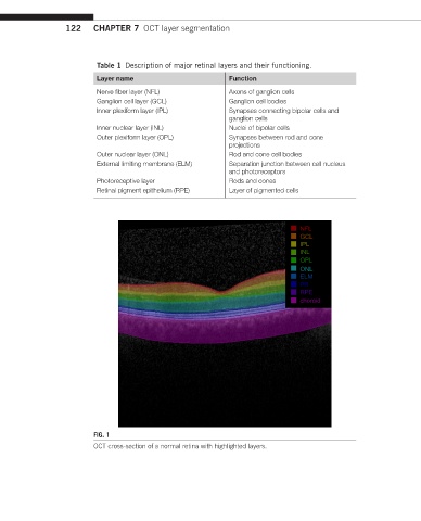


                                                                              NFL
                                                                              GCL
                                                                              IPL
                                                                              INL
                                                                              OPL
                                                                              ONL
                                                                              ELM
                                                                              PR
                                                                              RPE
                                                                              choroid


                         FIG. 1
                         OCT cross-section of a normal retina with highlighted layers.
   124   125   126   127   128   129   130   131   132   133   134