
Page 130 - Computational Retinal Image Analysis
P. 130
2 Algorithmic evaluation and benchmarking 123
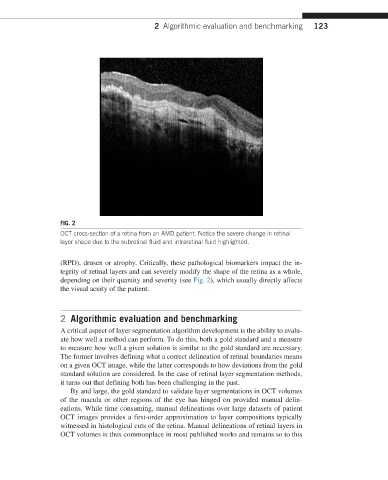
FIG. 2
OCT cross-section of a retina from an AMD patient. Notice the severe change in retinal
layer shape due to the subretinal fluid and intraretinal fluid highlighted.
(RPD), drusen or atrophy. Critically, these pathological biomarkers impact the in-
tegrity of retinal layers and can severely modify the shape of the retina as a whole,
depending on their quantity and severity (see Fig. 2), which usually directly affects
the visual acuity of the patient.
2 Algorithmic evaluation and benchmarking
A critical aspect of layer segmentation algorithm development is the ability to evalu-
ate how well a method can perform. To do this, both a gold standard and a measure
to measure how well a given solution is similar to the gold standard are necessary.
The former involves defining what a correct delineation of retinal boundaries means
on a given OCT image, while the latter corresponds to how deviations from the gold
standard solution are considered. In the case of retinal layer segmentation methods,
it turns out that defining both has been challenging in the past.
By and large, the gold standard to validate layer segmentations in OCT volumes
of the macula or other regions of the eye has hinged on provided manual delin-
eations. While time consuming, manual delineations over large datasets of patient
OCT images provides a first-order approximation to layer compositions typically
witnessed in histological cuts of the retina. Manual delineations of retinal layers in
OCT volumes is thus commonplace in most published works and remains so to this