
Page 135 - Computational Retinal Image Analysis
P. 135
128 CHAPTER 7 OCT layer segmentation
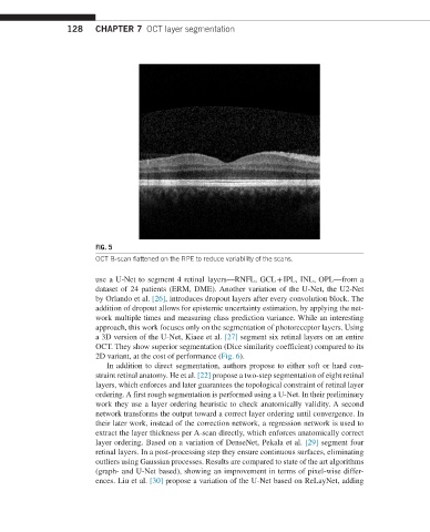
FIG. 5
OCT B-scan flattened on the RPE to reduce variability of the scans.
use a U-Net to segment 4 retinal layers—RNFL, GCL + IPL, INL, OPL—from a
dataset of 24 patients (ERM, DME). Another variation of the U-Net, the U2-Net
by Orlando et al. [26], introduces dropout layers after every convolution block. The
addition of dropout allows for epistemic uncertainty estimation, by applying the net-
work multiple times and measuring class prediction variance. While an interesting
approach, this work focuses only on the segmentation of photoreceptor layers. Using
a 3D version of the U-Net, Kiaee et al. [27] segment six retinal layers on an entire
OCT. They show superior segmentation (Dice similarity coefficient) compared to its
2D variant, at the cost of performance (Fig. 6).
In addition to direct segmentation, authors propose to either soft or hard con-
straint retinal anatomy. He et al. [22] propose a two-step segmentation of eight retinal
layers, which enforces and later guarantees the topological constraint of retinal layer
ordering. A first rough segmentation is performed using a U-Net. In their preliminary
work they use a layer ordering heuristic to check anatomically validity. A second
network transforms the output toward a correct layer ordering until convergence. In
their later work, instead of the correction network, a regression network is used to
extract the layer thickness per A-scan directly, which enforces anatomically correct
layer ordering. Based on a variation of DenseNet, Pekala et al. [29] segment four
retinal layers. In a post-processing step they ensure continuous surfaces, eliminating
outliers using Gaussian processes. Results are compared to state of the art algorithms
(graph- and U-Net based), showing an improvement in terms of pixel-wise differ-
ences. Liu et al. [30] propose a variation of the U-Net based on ReLayNet, adding