
Page 26 - Academic Press Encyclopedia of Physical Science and Technology 3rd BioChemistry
P. 26
P1: ZCK Final Pages
Encyclopedia of Physical Science and Technology EN005G-231 June 15, 2001 20:46
Enzyme Mechanisms 633
Measurement of this step in the reverse direction (i.e., DHFR protein molecule. The protein fold through its com-
for DHFR · NADP · H 4 F) coupled with determination of plex vibrational modes apparently may couple some set
+
the overall equilibrium constant permitted construction of of motions to a promotional vibration that fosters passage
Fig. 6. of the reactive ternary complex over the activation barrier.
The kinetic scheme served as the basis for the explana-
tion of the contribution of various elements of the protein
C. Phosphate Transfer
to its function. Site-specific mutagenesis is a technique
in which one or more amino acids are replaced by other Enzymes that catalyze the transfer of a phosphoryl moiety
amino acids through alteration of the gene encoding the between two substrates have provided excellent examples
enzyme. For the mutant proteins, the same kinetic scheme of the use of isotopes in kinetic and stereochemical stud-
was reconstructed to calculate the free energy differences ies. The enzyme hexokinase, which promotes the con-
arising from changes in the kinetic steps caused by the mu- version of glucose plus ATP to glucose-6-phosphate and
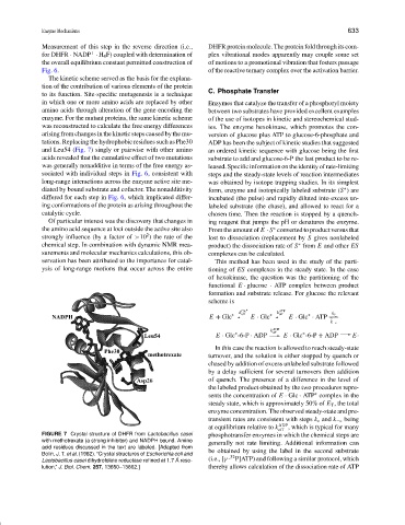
tations. Replacing the hydrophobic residues such as Phe30 ADP has been the subject of kinetic studies that suggested
and Leu54 (Fig. 7) singly or pairwise with other amino an ordered kinetic sequence with glucose being the first
acids revealed that the cumulative effect of two mutations substrate to add and glucose-6-P the last product to be re-
was generally nonadditive in terms of the free energy as- leased.Specificinformationontheidentityofrate-limiting
sociated with individual steps in Fig. 6, consistent with steps and the steady-state levels of reaction intermediates
long-range interactions across the enzyme active site me- was obtained by isotope trapping studies. In its simplest
diated by bound substrate and cofactor. The nonadditivity form, enzyme and isotopically labeled substrate (S ) are
∗
differed for each step in Fig. 6, which implicated differ- incubated (the pulse) and rapidly diluted into excess un-
ing conformations of the protein as arising throughout the labeled substrate (the chase), and allowed to react for a
catalytic cycle. chosen time. Then the reaction is stopped by a quench-
Of particular interest was the discovery that changes in ing reagent that jumps the pH or denatures the enzyme.
the amino acid sequence at loci outside the active site also From the amount of E · S converted to product versus that
∗
2
strongly influence (by a factor of >10 ) the rate of the lost to dissociation (replacement by S gives nonlabeled
chemical step. In combination with dynamic NMR mea- product) the dissociation rate of S from E and other ES
∗
surements and molecular mechanics calculations, this ob- complexes can be calculated.
servation has been attributed to the importance for catal- This method has been used in the study of the parti-
ysis of long-range motions that occur across the entire tioning of ES complexes in the steady state. In the case
of hexokinase, the question was the partitioning of the
functional E · glucose · ATP complex between product
formation and substrate release. For glucose the relevant
scheme is
k Glc ∗ k ATP k c
E + Glc ∗ off E · Glc ∗ off E · Glc · ATP
∗
k −c
k ADP
E · Glc -6-P · ADP off E · Glc -6-P + ADP E·
∗
∗
In this case the reaction is allowed to reach steady-state
turnover, and the solution is either stopped by quench or
chased by addition of excess unlabeled substrate followed
by a delay sufficient for several turnovers then addition
of quench. The presence of a difference in the level of
the labeled product obtained by the two procedures repre-
∗
sents the concentration of E · Glc · ATP complex in the
steady state, which is approximately 50% of E T , the total
enzyme concentration. The observed steady-state and pre-
transient rates are consistent with steps k c and k −c being
at equilibrium relative to k ADP , which is typical for many
off
FIGURE 7 Crystal structure of DHFR from Lactobacillus casei phosphotransfer enzymes in which the chemical steps are
with methotrexate (a strong inhibitor) and NADPH bound. Amino generally not rate limiting. Additional information can
acid residues discussed in the text are labeled. [Adapted from
Bolin, J. T. et al. (1982). “Crystal structures of Escherichia coli and be obtained by using the label in the second substrate
32
Lactobacillus casei dihydrofolate reductase refined at 1.7 ˚ A reso- (i.e., [γ - P]ATP) and following a similar protocol, which
lution,” J. Biol. Chem. 257, 13650–13662.] thereby allows calculation of the dissociation rate of ATP