
Page 260 - Fundamentals of Light Microscopy and Electronic Imaging
P. 260
VIDEO ENHANCEMENT OF IMAGE CONTRAST 243
al., 1981a,b; Inoué, 1981). As discussed in the previous chapter and shown in Figure
12-10, increasing the gain amplifies the signal, while offset is used to add or subtract a
voltage from the signal to bring background features close to the black level of the
image. In this way, a very narrow range of the video signal can be stretched to fill the
available gray levels ranging from black to white. Frame averaging, background sub-
traction, and further contrast adjustments are made using an in-line digital image
processor to produce a high-contrast, high-quality image. This procedure, known as
video enhancement of image contrast, was introduced and popularized by Shinya Inoué,
Robert Allen, and others, and has been used to view objects such as lamellapodia and
endoplasmic reticulum in living cells. The technique is so sensitive that it is possible to
image purified microtubules, which have been shown to retard a wavelength of green
light by only /300 (Fig. 13-8)! The procedure for performing video enhancement of
image contrast is as follows:
(a)
(b)
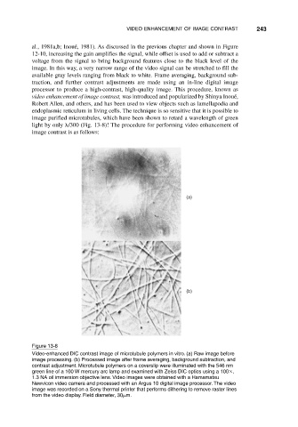
Figure 13-8
Video-enhanced DIC contrast image of microtubule polymers in vitro. (a) Raw image before
image processing. (b) Processed image after frame averaging, background subtraction, and
contrast adjustment. Microtubule polymers on a coverslip were illuminated with the 546 nm
green line of a 100 W mercury arc lamp and examined with Zeiss DIC optics using a 100 ,
1.3 NA oil immersion objective lens. Video images were obtained with a Hamamatsu
Newvicon video camera and processed with an Argus 10 digital image processor. The video
image was recorded on a Sony thermal printer that performs dithering to remove raster lines
from the video display. Field diameter, 30 m.