
Page 268 - Handbook of Deep Learning in Biomedical Engineering Techniques and Applications
P. 268
Chapter 9 Applications of deep learning in biomedical engineering 259
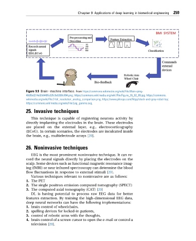
Figure 9.9 Brainemachine interface. From https://commons.wikimedia.org/wiki/File:Main-qimg-
48d5bd214e53d440fa32fc9e5300c894.png; https://commons.wikimedia.org/wiki/File:Figure_35_02_09.jpg; https://commons.
wikimedia.org/wiki/File:2-bit_resolution_analog_comparison.png; https://www.pikrepo.com/fktpp/black-and-gray-robot-toy;
https://commons.wikimedia.org/wiki/File:Eeg_gamma.svg.
25. Invasive techniques
This technique is capable of registering neurons activity by
directly implanting the electrodes in the brain. These electrodes
are placed on the external layer, e.g., electrocorticography
(ECoG). In certain scenarios, the electrodes are inculcated inside
the brain, e.g., multielectrode arrays [20].
26. Noninvasive techniques
EEG is the most prominent noninvasive technique. It can re-
cord the neural signals directly by placing the electrodes on the
scalp. Some devices such as functional magnetic resonance imag-
ing (fMRI) or near infrared spectroscopy can determine the blood
flow fluctuations in response to external stimuli [20].
Various techniques relevant to noninvasive are as follows:
1. The PET
2. The single positron emission computed tomography (SPECT)
3. The computed axial tomography (CAT) [20]
DL is having potential to process raw EEG data for better
features extraction. By training the high-dimensional EEG data,
deep neural networks can have the following implementations:
1. brain control of wheelchairs,
2. spelling devices for locked-in patients,
3. control of robotic arms with the thoughts,
4. brain control of a screen cursor to open the e-mail or control a
television [20].