
Page 224 - Vibrational Spectroscopic Imaging for Biomedical Applications
P. 224
200 Cha pte r Se v e n
Cornea Retina
Lens
(a)
~ 4 mm
(b)
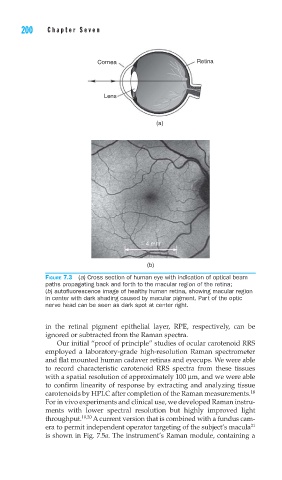
FIGURE 7.3 (a) Cross section of human eye with indication of optical beam
paths propagating back and forth to the macular region of the retina;
(b) autofl uorescence image of healthy human retina, showing macular region
in center with dark shading caused by macular pigment. Part of the optic
nerve head can be seen as dark spot at center right.
in the retinal pigment epithelial layer, RPE, respectively, can be
ignored or subtracted from the Raman spectra.
Our initial “proof of principle” studies of ocular carotenoid RRS
employed a laboratory-grade high-resolution Raman spectrometer
and flat mounted human cadaver retinas and eyecups. We were able
to record characteristic carotenoid RRS spectra from these tissues
with a spatial resolution of approximately 100 μm, and we were able
to confirm linearity of response by extracting and analyzing tissue
18
carotenoids by HPLC after completion of the Raman measurements.
For in vivo experiments and clinical use, we developed Raman instru-
ments with lower spectral resolution but highly improved light
throughput. 19,20 A current version that is combined with a fundus cam-
21
era to permit independent operator targeting of the subject’s macula
is shown in Fig. 7.5a. The instrument’s Raman module, containing a