
Page 228 - Vibrational Spectroscopic Imaging for Biomedical Applications
P. 228
204 Cha pte r Se v e n
2500
Macular pigments, M ± S.D. 2000
1500
1000
500
0
22 24 26 28
Age, yrs
(a)
Macular pigments, M ± S.D. 2500
2000
1500
1000
500
0
20 30 40 50 60 70 80 90
Age, yrs
(b)
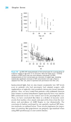
FIGURE 7.6 (a) RRS MP measurements of 33 normal eyes for a young group of
subjects ranging in age from 21 to 29 years. Note the large (up to ~10-fold)
variation of RRS levels that can exist between individuals; (b) RRS
measurements of 212 normal eyes as a function of subject age, revealing
statistically signifi cant decrease of MP concentration with age. (Panel a
adapted from Ref. 26, panel b reprinted with permission from Ref. 20.)
backscattered light, but we also found consistently low MP levels
even in patients who had previously had cataract surgery with
implantation of optically clear prosthetic intraocular lenses (pseudo-
phakia). Also, we have noted that patients with unilateral cataracts
after trauma or retinal detachment repair typically have very similar
RRS carotenoid levels in the normal and in the pseudophakic eye.
Thus, we have concluded that there is a decline of macular carot-
enoids that reaches a low steady state just at the time when the inci-
dence and prevalence of AMD begins to rise dramatically. The
conclusion is further confirmed by our spatially resolved MP detec-
tion methods, as outlined below. These results emphasize the impor-
tance of assuring that populations are properly age-matched when
using RRS spectroscopy in case-control studies.