Page 232 - Vibrational Spectroscopic Imaging for Biomedical Applications
P. 232
208    Cha pte r  Se v e n


            The first term describes fluorescence from the ocular media (OM),
        the second term the Raman response from macular pigment (MP),
        and the third term fluorescence from lipofuscin (LP). In Eq. (7.1), α
                                                                OM
        and α  are the absorption coefficients of the optical media and lipo-
             LP
        fuscin, respectively; T   and T  are the percentage transmissions of
                           OM     MP
        the optical media and MP at the indicated absorption and emission
        wavelengths;  N MP  is the concentration of the MP molecules,  σ MP
        their Raman scattering cross section, and η   and η  describe the
                                              OM     LP
        quantum efficiencies for optical media and lipofuscin fluorescence
        transitions.
            The detector signal at the Raman wavelength is a superposition
        of a weak, spectrally narrow Raman signal, and typically 100-fold
        stronger and spectrally broad background fluorescence (mostly from
        the lens and lipofuscin). To retrieve the MP Raman response, the
        superimposed fluorescence intensities need to be measured and sub-
        tracted from the total detector signal. The contribution of the broad
        fluorescence at the Raman wavelength is approximately the same as
        at a wavelength slightly offset to a longer wavelength position, λ  .
                                                               offset
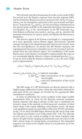
        It can be shown that the Raman component, I (λ ), for each image
                                                R  R
        pixel is approximately
              I (λ  ) ≈  T −1  (λ  )  T ⋅  −1  (λ  I )(  (λ  )/ T −  I  (λ  / )(T  )
             R  R    OM  exc  OM  R  Det  R  R  Det  o offset  offset

        where I  (λ  ) and I  (λ  ) = detector intensities
               Det  R    Det  offset
                       T  and T   =  filter transmissions at the respective
                        R     offset
                                   wavelengths
                             T    =  unknown transmission of the ocular
                              OM
                                   media
            The RRI image of a MP distribution can thus be derived with a
        digital image subtraction routine, where the intensities obtained for
        each pixel of the two images are divided by the appropriate filter
        transmission coefficient.
            In order to determine the correct position of the angle tunable
        transmission filter, to check for the linearity between resonance
        Raman response and optical density of MP, and to determine the
        dynamic range of the method, we first measured a number of dried
        lutein drops with widely differing concentration levels.  All drops
        were spotted onto polyvinyl pyrolidine difluoride (PVDF) substrates.
        In Fig. 7.9 we show the RRI image obtained for the lutein drop with
        the highest concentration level in the center (OD ≈ 0.8). The intensity
        of each pixel is color coded according to the linear intensity scale
        shown at right. Similar imaging measurements were carried out with
        half a dozen drops of varying lutein concentrations. The integrated
        response was found to be roughly linear in concentration up to the
        highest level.

