
Page 231 - Vibrational Spectroscopic Imaging for Biomedical Applications
P. 231
Raman Detection of Car otenoids in Human T issue 207
2
2
the retinal radiant exposure is (4 mW)(400 ms)/0.096 cm = 16.7 mJ/cm ,
assuming all radiant power enters the pupil. This level is considered
25
safe according to limits set by the ANSI standard. The laser light
exposures caused after-images that typically disappeared within a
few minutes. During this time, the filters were changed to switch
from Raman to fluorescence imaging.
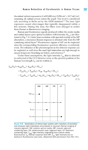
Raman and fluorescence signals produced within the ocular media
and retinal layers upon optical excitation with intensity I(λ ) are illus-
exc
trated in Fig. 7. 8. Under laser excitation with spectral overlap of the MP
absorption, a resonance Raman response is obtained only from the MP
26
containing retinal layer. Fluorescence signals of MP can be neglected
since the corresponding fluorescence quantum efficiency is extremely
weak. The influence of the photoreceptors in the detector response can
be neglected as well since the used light intensities are high enough to
ensure temporary bleaching see below, and reference. 27
Under these assumptions, the light intensity I that is detected
Det
in each pixel of the CCD detector array at the spectral position of the
Raman wavelength λ can be written as
R
⋅
=
I (λ ) α (λ ) η (λ )⋅ I(λ )
Det R OM exc OM R exc
⋅
+ T (λ )⋅T (λ )⋅ N ⋅σ (λ ) (λ )
I
OM exc OM R MP MP R exc
+ T (λ )⋅T (λ )⋅α (λ )⋅ η (λ )(⋅ λ )⋅ T (λ ) T ⋅ (λ )
I
OM exc MP exc LP exc LP R R exc MP R OM R
(7.1)
OM MP PR LP
I OM
I Exc
I R
I LP
FIGURE 7.8 Schematics of retinal layer system with indication of excitation
and emission light intensities encountered in resonant Raman imaging of
macular pigment. OM: anterior optical media, MP: macular pigment
containing layer; OS: photoreceptor outer segment layer; LP: lipofuscin
containing layer. I OM : light intensity originating from optical media
fl uorescence; I : Raman response from MP; I : light intensity due to
R LP
lipofuscin fl uorescence (see text). (Reprinted with permission from Ref. 24.)