
Page 212 - Bio Engineering Approaches to Cancer Diagnosis and Treatment
P. 212
8.5 Application of HIFU on thermal ablation 211
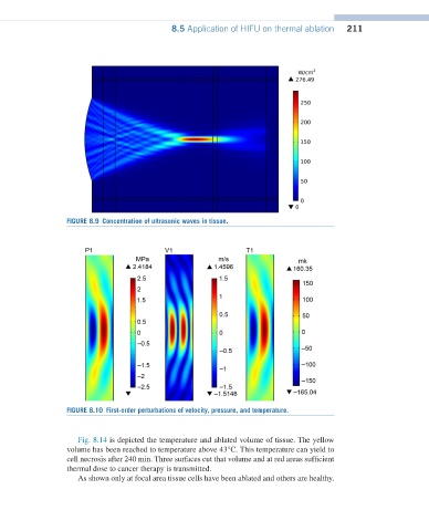
FIGURE 8.9 Concentration of ultrasonic waves in tissue.
FIGURE 8.10 First-order perturbations of velocity, pressure, and temperature.
Fig. 8.14 is depicted the temperature and ablated volume of tissue. The yellow
volume has been reached to temperature above 43°C. This temperature can yield to
cell necrosis after 240 min. Three surfaces cut that volume and at red areas sufficient
thermal dose to cancer therapy is transmitted.
As shown only at focal area tissue cells have been ablated and others are healthy.