
Page 147 - Biomedical Engineering and Design Handbook Volume 2, Applications
P. 147
126 MEDICAL DEVICE DESIGN
Needle
High vacuum
valve
pump
A B C
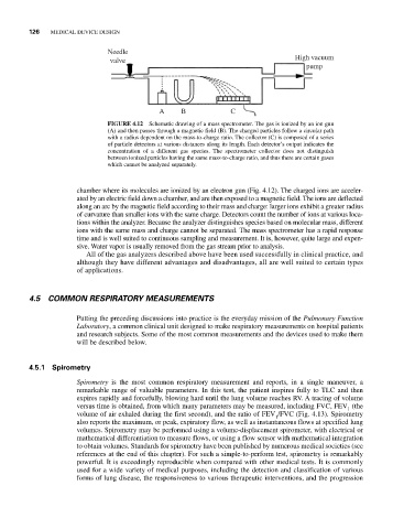
FIGURE 4.12 Schematic drawing of a mass spectrometer. The gas is ionized by an ion gun
(A) and then passes through a magnetic field (B). The charged particles follow a circular path
with a radius dependent on the mass-to-charge ratio. The collector (C) is composed of a series
of particle detectors at various distances along its length. Each detector’s output indicates the
concentration of a different gas species. The spectrometer collector does not distinguish
between ionized particles having the same mass-to-charge ratio, and thus there are certain gases
which cannot be analyzed separately.
chamber where its molecules are ionized by an electron gun (Fig. 4.12). The charged ions are acceler-
ated by an electric field down a chamber, and are then exposed to a magnetic field. The ions are deflected
along an arc by the magnetic field according to their mass and charge: larger ions exhibit a greater radius
of curvature than smaller ions with the same charge. Detectors count the number of ions at various loca-
tions within the analyzer. Because the analyzer distinguishes species based on molecular mass, different
ions with the same mass and charge cannot be separated. The mass spectrometer has a rapid response
time and is well suited to continuous sampling and measurement. It is, however, quite large and expen-
sive. Water vapor is usually removed from the gas stream prior to analysis.
All of the gas analyzers described above have been used successfully in clinical practice, and
although they have different advantages and disadvantages, all are well suited to certain types
of applications.
4.5 COMMON RESPIRATORY MEASUREMENTS
Putting the preceding discussions into practice is the everyday mission of the Pulmonary Function
Laboratory, a common clinical unit designed to make respiratory measurements on hospital patients
and research subjects. Some of the most common measurements and the devices used to make them
will be described below.
4.5.1 Spirometry
Spirometry is the most common respiratory measurement and reports, in a single maneuver, a
remarkable range of valuable parameters. In this test, the patient inspires fully to TLC and then
expires rapidly and forcefully, blowing hard until the lung volume reaches RV. A tracing of volume
versus time is obtained, from which many parameters may be measured, including FVC, FEV (the
1
volume of air exhaled during the first second), and the ratio of FEV /FVC (Fig. 4.13). Spirometry
1
also reports the maximum, or peak, expiratory flow, as well as instantaneous flows at specified lung
volumes. Spirometry may be performed using a volume-displacement spirometer, with electrical or
mathematical differentiation to measure flows, or using a flow sensor with mathematical integration
to obtain volumes. Standards for spirometry have been published by numerous medical societies (see
references at the end of this chapter). For such a simple-to-perform test, spirometry is remarkably
powerful. It is exceedingly reproducible when compared with other medical tests. It is commonly
used for a wide variety of medical purposes, including the detection and classification of various
forms of lung disease, the responsiveness to various therapeutic interventions, and the progression