
Page 149 - Biomedical Engineering and Design Handbook Volume 2, Applications
P. 149
128 MEDICAL DEVICE DESIGN
6
MP BV 5 Inspiration
Volume (L) 3 collection
Analyzers 4 Alveolar
SB sample
2
Breath-hold Washout
1
0
0 2 4 6 8 10 12 14 16
Time (s)
A B
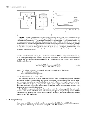
FIGURE 4.14 Illustration of equipment for performing a single-breath diffusing capacity test (a). The patient breathes
through the mouthpiece (MP). Initially, the breathing valve (BV) is turned so that all breathing is in from and out to the
room. After the patient exhales to RV, the breathing valve is turned to attach the patient to the spirometer filled with test
gas containing carbon monoxide and helium. The patient inspires rapidly to TLC, breath-holds for 10 seconds, and
exhales rapidly. The breathing valve is left connected to the spirometer for the initial portion of this expiration, allowing
the spirometer to record the amount of gas washing out the dead space. After the dead space has been flushed, the breath-
ing valve is turned so tht an alveolar sample is collected in the sample bag (SB). Gas analyzers measure the inspired gas
concentrations from the spirometer, and the expired gas concentrations from the sample bag. A representative spirome-
ter tracing is shown on the right (b).
Over the period of breath holding, the alveolar concentration of CO falls exponentially according
to its partial pressure gradient between the gas and blood sides of the alveolar membrane (it is
assumed that the blood concentration of CO is zero throughout the short breath-hold). Then, the
DLCO is calculated as
TR 1 1 CO ⎞
F I ⎛ F A
DLCO = V I ln ⎜ ⎟ ⎠ (4.10)
TR T BP − 47 ⎝ F E
F E CO
where V = volume of inspired gas (usually adjusted by an estimate of dead space)
I
T = duration of breath-hold
BP = ambient barometric pressure
and other parameters are as defined above.
The equipment needed to calculate the DLCO includes either a spirometer or a flow sensor to
measure the inspired volume and gas analyzers to measure the concentrations of CO and the tracer
gas. In some systems, the sample of expired alveolar gas is collected in a bag for analysis; in other
systems with rapidly responding analyzers, the expired gas is sampled continuously for calculation.
It is worth noting that using a flow sensor for this test requires that the flow sensor be calibrated with
the gases to be used, as described above.
The DLCO is very sensitive for lung abnormalities but is also quite nonspecific. Several condi-
tions can cause marked reductions in DLCO, including emphysema and pulmonary fibrosis. DLCO
can be increased in some cases of early heart failure and in cases of pulmonary hemorrhage. DLCO
is reported at STPD conditions.
4.5.3 Lung Volumes
There are several different methods available for measuring the TLC, RV, and FRC. These parame-
ters, regardless of how they are measured, are reported at BTPS conditions.