
Page 151 - Biomedical Engineering and Design Handbook Volume 2, Applications
P. 151
130 MEDICAL DEVICE DESIGN
Flowmeter
A A
100%
O 2 Nitrogen
analyzer
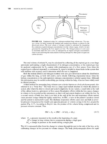
FIGURE 4.16 Equipment setup for a nitrogen-washout lung volume test. The one-
way valves (A) keep separate the inspired and expired gas streams, as shown by the
directional arrows. The total volume of nitrogen exhaled is calculated by integrating
the continuous expired nitrogen concentration over the volume signal, which is in turn
generated by integrating the flow signal over time. The nitrogen analyzer is also used
to determine when the test is over, as indicated by an expired nitrogen concentration
close to zero following the replacement of all lung nitrogen by other gases (oxygen and
carbon dioxide).
The total volume of exhaled N may be calculated by collecting all the expired gas in a very large
2
spirometer and making a single measurement of its nitrogen concentration, or the expired gas may
be analyzed continuously for N content with simultaneous use of a flow sensor. Even when the
2
expired gas is collected separately, a continuous N signal is helpful to detect leaks in the system that
2
will interfere with accuracy and to determine when the test is completed.
Both the helium-dilution and nitrogen-washout tests also give information about the distribution
of gas within the lung, as both will yield a curve closely following exponential decay when the
helium concentration or the end-expiratory nitrogen concentration is plotted against time. Although
this information may be useful in describing gas mixing within the lung, it has not been widely used
in clinical practice.
A third method for measuring lung volumes uses a device known as a body plethysmograph, also
referred to as a “body box.” The body box is a large rigid-walled structure in which the patient is
seated, after which the door is closed and sealed completely. In one variety, a small hole in the wall
of the cabinet leads to a spirometer or flow sensor. Respiratory efforts within the box causes changes
in volume to be recorded on this spirometer as chest wall movement displaces air within the box. In
a second variety of body box, there is no hole in the wall of the box and respiratory efforts instead
cause pressure swings within the tightly sealed box. At FRC, a valve at the patient’s mouth is closed
and the patient is instructed to pant. The rhythmic respiratory efforts cause rises and falls in alveo-
lar pressure (measured at the mouth) and opposite pressure or volume swings in the box around the
patient (Fig. 4.17). According to Boyle’s law, the product of the volume being compressed and its
pressure remains constant. Thus
Δ
Δ
)(
FRC × P m = ( FRC − V P m + P m ) (4.13)
where P = pressure measured at the mouth at the beginning of a pant
m
ΔV = change in lung volume due to compression during a single pant
ΔP = change in pressure at the mouth during a single pant
m
The ΔV is measured either from the change in volume through the port in the side of the box, or by
calibrating changes in box pressure to volume changes. The body plethysmograph allows for rapid