
Page 303 - Color Atlas of Biochemistry
P. 303
294 Tissues and organs
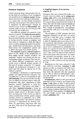
Immune response A. Simplified diagram of the immune
response
Viruses, bacteria, fungi, and parasites that en-
ter the body of vertebrates of are recognized Pathogens that have entered the body—e. g.,
viruses (top)—are taken up by antigen-pre-
and attacked by the immune system.Endog-
enous cells that have undergone alterations— senting cells (APCs) and proteolytically de-
graded (1). The viral fragments produced in
e. g., tumor cells—are also usually recognized
as foreign and destroyed. The immune system this way are then presented on the surfaces of
these cells with the help of special membrane
is supported by physiological changes in
infected tissue, known as inflammation.This proteins (MHC proteins; see p. 296) (2). The
reaction makes it easier for the immune cells APCs include B lymphocytes, macrophages,
and dendritic cells such as the skin’s Langer-
to reach the site of infection.
Two different systems are involved in the hans cells.
immune response. The innate immune system The complexes of MHC proteins and viral
is based on receptors that can distinguish fragments displayed on the APCs are recog-
between bacterial and viral surface structures nized by T cells that carry a receptor that
matches the antigen (“T-cell receptors”; see
or foreign proteins (known as antigens)and
those that are endogenous. With the help of p. 296) (3). Binding leads to activation of the T
these receptors, phagocytes bind to the patho- cell concerned and selective replication of it
gens, absorb them by endocytosis, and break (4, “clonal selection”). The proliferation of im-
them down. The complement system (see mune cells is stimulated by interleukins (IL).
These are a group of more than 20 signaling
p. 298) is also part of the innate system.
The acquired (adaptive) immune system is substances belonging to the cytokine family
based on the ability of the lymphocytes to (see p. 392), with the help of which immune
form highly specific antigen receptors “on cells communicate with each other. For exam-
suspicion,” without ever having met the cor- ple, activated macrophages release IL-1 (5),
while T cells stimulate their own replication
responding antigen. In humans, there are sev-
eral billion different lymphocytes, each of and that of other immune cells by releasing
IL-2 (6).
whichcarries adifferent antigenreceptor. If
this type of receptor recognizes “its” cognate Depending on their type, activated T cells
antigen, the lymphocyte carrying it is acti- have different functions. Cytotoxic T cells
(green) are able to recognize and bind virus-
vated and then plays its special role in the
immune response. infected body cells or tumor cells (7). They
then drive the infected cells into apoptosis
In addition, a distinction is made between
cellular and humoral immune responses. The (see p. 396) or kill them with perforin, apro-
Tlymphocytes (T cells) are responsible for cel- tein that perforates the target cell’s plasma
membrane (8).
lular immunity. They are named after the thy-
B lymphocytes, which as APCs present viral
mus, in which the decisive steps in their dif- fragments on their surfaces, are recognized by
ferentiation take place. Depending on their
function, another distinction is made be- helper T cells (blue) or their T cell receptors
tween cytotoxic T cells (green) and helper T (9). Stimulated by interleukins, selective clo-
nal replication then takes place of B cells that
cells (blue). Humoral immunity is based on
the activity of the B lymphocytes (B cells, light carry antigen receptors matching those of the
brown), which mature in the bone marrow. pathogen (10). Thesematureinto plasma cells
After activation by T cells, B cells are able to (11) and finally secrete large amounts of
release soluble forms of their specific antigen soluble antibodies (12).
receptors, known as antibodies (see p. 300),
into the blood plasma. The immune system’s
“memory” is represented by memory cells.
These are particularly long–lived cells that
can arise from any of the lymphocyte types
described.
Koolman, Color Atlas of Biochemistry, 2nd edition © 2005 Thieme
All rights reserved. Usage subject to terms and conditions of license.