
Page 305 - Color Atlas of Biochemistry
P. 305
296 Tissues and organs
T-cell activation that any two individuals carry the same set of
MHC proteins—except for monozygotic twins.
For the selectivity of the immune response Class I MHC proteins occur in almost all
(see p. 294), the cells involved must be able nucleated cells. They mainly interact with cy-
to recognize foreign antigens and proteins on totoxic T cells and are the reason for the re-
other immune cells safely and reliably. To do jection of transplanted organs. Class I MHC
this, they have antigen receptors on their cell proteins are heterodimers (αβ). The β subunit
surfaces and co-receptors that support recog- is also known as β 2 -microglobulin.
nition. Class II MHC proteins also consist of two
peptide chains, which are related to each
other. MHC II molecules are found on all anti-
A. Antigen receptors
gen-presenting cells in the immune system.
Many antigen receptors belong to the immu- They serve for interaction between these cells
noglobulin superfamily. The common charac- and CD4-carrying T helper cells.
teristic of these proteins is that they are made
up from “immunoglobulin domains.” These
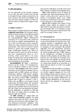
are characteristically folded substructures B. T-cell activation
consisting of 70–110 amino acids, which are The illustration shows an interaction between
also found in soluble immunoglobulins (Ig; a virus-infected body cell (bottom) and a CD8-
see p. 300). The illustration shows schemati- carrying cytotoxic T lymphocyte (top). The
cally a few of the important proteins in the Ig infected cell breaks down viral proteins in
superfamily. They consist of constant regions its cytoplasm (1) and transports the peptide
(brown or green) and variable regions (or- fragments into the endoplasmic reticulum
ange). Homologous domains are shown in with the help of a special transporter (TAP)
the same colors in each case. All of the recep- (2). Newly synthesized class I MHC proteins
tors have transmembrane helices at the C on the endoplasmic reticulum are loaded
terminus, which anchor them to the mem- with one of the peptides (3)and then trans-
branes. Intramolecular and intermolecular di- ferred to the cell surface by vesicular trans-
sulfide bonds are also usually found in pro- port (4). The viral peptides are bound on the
teins belonging to the Ig family. surface of the α 2 domain of the MHC protein
Immunoglobulin M (IgM), a membrane in a depressionformed by aninsertion as a
proteinonthe surface of B lymphocytes, “floor” and two helices as “walls” (see smaller
serves to bind free antigens to the B cells. By illustration).
contrast, T cell receptors only bind antigens Supported by CD8 and other co-receptors,
when they are presented by another cell as a a T cell with a matching T cell receptor binds
complexwithanMHC protein(see below). to the MHC peptide complex (5; cf. p. 224).
Interaction between MHC-bound antigens This binding activates protein kinases in the
and T cell receptors is supported by co-recep- interior of the T cell, which trigger a chain of
tors. This group includes CD8, a membrane additional reactions (signal transduction; see
protein that is typical in cytotoxic T cells. T p. 388). Finally, destruction of the virus-in-
helper cells use CD4 as a co-receptor instead fected cell by the cytotoxic T lymphocytes
(not shown). The abbreviation “CD” stands for takes place.
“cluster of differentiation.” It is the term for a
large group of proteins that are all located on
the cell surface and can therefore be identi-
fied by antibodies. In addition to CD4 and
CD8, there are many other co-receptors on
immune cells (not shown).
The MHC proteins are named after the
“major histocompatibility complex”—the DNA
segmentthatcodes for them. Human MHC
proteins are also known as HLA antigens (“hu-
man leukocyte-associated” antigens). Their
polymorphism is so large that it is unlikely
Koolman, Color Atlas of Biochemistry, 2nd edition © 2005 Thieme
All rights reserved. Usage subject to terms and conditions of license.