
Page 252 - Academic Press Encyclopedia of Physical Science and Technology 3rd Analytical Chemistry
P. 252
P1: GRB/GWT P2: GPJ/GAX QC: GAE/FYD Final Pages
Encyclopedia of Physical Science and Technology EN008M-395 June 29, 2001 15:52
Magnetic Resonance in Medicine 977
The total time required to complete a scan is M times
T R , so that T 2 -weighted images generally take longer to
acquire. Often it is desired to enhance the SNR by repeat-
ing the entire sequence one or more times and averaging
the results of corresponding cycles. If there are n repeti-
tions of the basic sequence, the total scan time increases to
√
nMT R and the SNR is increased by n. The total time to
complete an individual scan usually ranges from about 1
to 15 min. Because it is usually necessary to make more
than one series of images, the patient is normally in the
magnet from 15 to 90 min to complete a diagnostic study.
IV. CLINICAL APPLICATIONS OF
MAGNETIC RESONANCE IMAGING
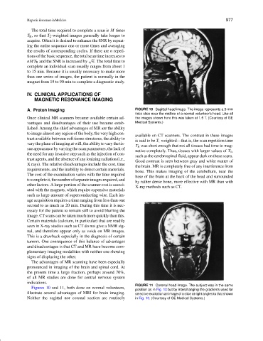
A. Proton Imaging FIGURE 10 Sagittal head image. The image represents a 3-mm
thick slice near the midline of a normal volunteer’s head. Like all
Once clinical MR scanners became available certain ad- the images shown here this was taken at 1.5 T. (Courtesy of GE
vantages and disadvantages of their use became estab- Medical Systems.)
lished. Among the chief advantages of MR are the ability
to image almost any region of the body, the very high con-
available on CT scanners. The contrast in these images
trast available between soft tissue structures, the ability to
is said to be T 1 weighted—that is, the scan repetition time
vary the plane of imaging at will, the ability to vary the tis-
T R was short enough that not all tissues had time to mag-
sue appearance by varying the scan parameters, the lack of
netize completely. Thus, tissues with larger values of T 1 ,
the need for any invasive step such as the injection of con-
such as the cerebrospinal fluid, appear dark on these scans.
trast agents, and the absence of any ionizing radiation (i.e.,
Good contrast is seen between gray and white matter of
X rays). The relative disadvantages include the cost, time
the brain. MR is completely free of any interference from
requirements, and the inability to detect certain materials.
bone. This makes imaging of the cerebellum, near the
The cost of the examination varies with the time required
base of the brain at the back of the head and surrounded
tocompleteit,thenumberofseparateimagesrequired,and
by rather dense bone, more effective with MR than with
other factors. A large portion of the scanner cost is associ-
X-ray methods such as CT.
ated with the magnets, which require expensive materials
such as large amount of superconducting wire. Each im-
age acquisition requires a time ranging from less than one
second to as much as 20 min. During this time it is nec-
essary for the patient to remain still to avoid blurring the
image.CTscanscanbetakenmuchmorequicklythanthis.
Certain materials (calcium, in particular) that are readily
seen in X-ray studies such as CT do not give a NMR sig-
nal, and therefore appear only as voids on MR images.
This is a drawback especially in the diagnosis of certain
tumors. One consequence of this balance of advantages
and disadvantages is that CT and MR have become com-
plementary imaging modalities with neither one showing
signs of displacing the other.
The advantages of MR scanning have been especially
pronounced in imaging of the brain and spinal cord. At
the present time a large fraction, perhaps around 70%,
of all MR studies are done for central nervous system
indications.
FIGURE 11 Coronal head image. The subject was in the same
Figures 10 and 11, both done on normal volunteers,
position as in Fig. 10 but by interchanging the gradients used for
illustrate several advantages of MRI for brain imaging. selective excitation an image of a slice at right angles to that shown
Neither the sagittal nor coronal section are routinely in Fig. 10. (Courtesy of GE Medical Systems.)