
Page 255 - Academic Press Encyclopedia of Physical Science and Technology 3rd Analytical Chemistry
P. 255
P1: GRB/GWT P2: GPJ/GAX QC: GAE/FYD Final Pages
Encyclopedia of Physical Science and Technology EN008M-395 June 29, 2001 15:52
980 Magnetic Resonance in Medicine
the standard platform for high performance clinical MRI.
In the late 1980s a number of research sites began to
make use of the improved signal-to-noise ratio available
at high field strengths by experimenting with whole-body
scanners operating at 4 T. By the end of the 1990s a
substantial clinical market began to develop for whole-
body clinical scanners operated at fields well above 1.5 T
—particularly at fields of 3 and 4 T. This trend was driven
initially by the interest of the neuroscience community
in blood-oxygen-level-dependent contrast (BOLD) func-
tional MRI (fMRI). This contrast mechanism is associ-
ated with the magnetic susceptibility difference between
oxygenated and deoxygenated hemoglobin in the cerebral
microvasculature, and susceptibility-based contrast is in-
herently greater at high field strength. The technique of
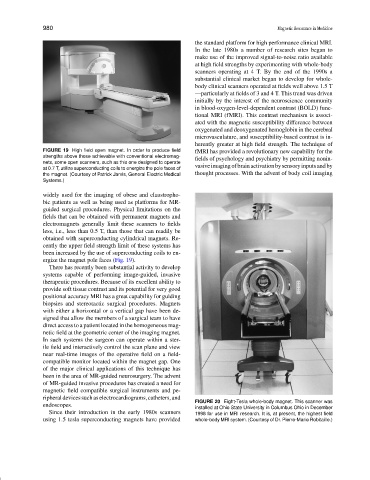
FIGURE 19 High field open magnet. In order to produce field fMRI has provided a revolutionary new capability for the
strengths above those achievable with conventional electromag-
fields of psychology and psychiatry by permitting nonin-
nets, some open scanners, such as this one designed to operate
vasiveimagingofbrainactivationbysensoryinputsandby
at 0.7 T, utilize superconducting coils to energize the pole faces of
the magnet. (Courtesy of Patrick Jarvis, General Electric Medical thought processes. With the advent of body coil imaging
Systems.)
widely used for the imaging of obese and claustropho-
bic patients as well as being used as platforms for MR-
guided surgical procedures. Physical limitations on the
fields that can be obtained with permanent magnets and
electromagnets generally limit these scanners to fields
less, i.e., less than 0.5 T, than those that can readily be
obtained with superconducting cylindrical magnets. Re-
cently the upper field strength limit of these systems has
been increased by the use of superconducting coils to en-
ergize the magnet pole faces (Fig. 19).
There has recently been substantial activity to develop
systems capable of performing image-guided, invasive
therapeutic procedures. Because of its excellent ability to
provide soft tissue contrast and its potential for very good
positional accuracy MRI has a great capability for guiding
biopsies and stereotactic surgical procedures. Magnets
with either a horizontal or a vertical gap have been de-
signed that allow the members of a surgical team to have
direct access to a patient located in the homogeneous mag-
netic field at the geometric center of the imaging magnet.
In such systems the surgeon can operate within a ster-
ile field and interactively control the scan plane and view
near real-time images of the operative field on a field-
compatible monitor located within the magnet gap. One
of the major clinical applications of this technique has
been in the area of MR-guided neurosurgery. The advent
of MR-guided invasive procedures has created a need for
magnetic field compatible surgical instruments and pe-
ripheraldevicessuchaselectrocardiograms,catheters,and
FIGURE 20 Eight-Tesla whole-body magnet. This scanner was
endoscopes.
installed at Ohio State University in Columbus Ohio in December
Since their introduction in the early 1980s scanners 1998 for use in MRI research. It is, at present, the highest field
using 1.5 tesla superconducting magnets have provided whole-body MRI system. (Courtesy of Dr. Pierre-Marie Robitaille.)