
Page 253 - Academic Press Encyclopedia of Physical Science and Technology 3rd Analytical Chemistry
P. 253
P1: GRB/GWT P2: GPJ/GAX QC: GAE/FYD Final Pages
Encyclopedia of Physical Science and Technology EN008M-395 June 29, 2001 15:52
978 Magnetic Resonance in Medicine
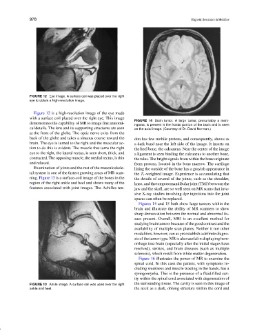
FIGURE 12 Eye image. A surface coil was placed over the right
eye to obtain a high-resolution image.
Figure 12 is a high-resolution image of the eye made
with a surface coil placed over the right eye. This image
FIGURE 14 Brain tumor. A large tumor, presumably a meni-
demonstrates the capability of MR to image fine anatomi-
ngoma, is present in the frontal portion of the brain and is seen
cal details. The lens and its supporting structures are seen on the axial image. (Courtesy of Dr. David Norman.)
at the front of the globe. The optic nerve exits from the
back of the globe and takes a sinuous course toward the don has few mobile protons, and consequently, shows as
brain. The eye is turned to the right and the muscular ac- a dark band near the left side of the image. It inserts on
tion to do this is evident. The muscle that turns the right the heel bone, the calcaneus. Near the center of the image
eye to the right, the lateral rectus, is seen short, thick, and a ligament is seen binding the calcaneus to another bone,
contracted. The opposing muscle, the medial rectus, is thin the talus. The bright signals from within the bone originate
and relaxed. from protons, located in the bone marrow. The cartilage
Examination of joints and the rest of the musculoskele- lining the outside of the bone has a grayish appearance in
tal system is one of the fastest growing areas of MR scan- the T 1 -weighted image. Experience is accumulating that
ning. Figure 13 is a surface-coil image of the bones in the the details of several of the joints, such as the shoulder,
region of the right ankle and heel and shows many of the knee, and the temporomandibular joint (TMJ) between the
features associated with joint images. The Achilles ten- jaw and the skull, are so well seen on MR scans that inva-
sive X-ray studies involving dye injections into the joint
spaces can often be replaced.
Figures 14 and 15 both show large tumors within the
brain and illustrate the ability of MR scanners to show
sharp demarcation between the normal and abnormal tis-
sues present. Overall, MRI is an excellent method for
studying brain tumors because of the good contrast and the
availability of multiple scan planes. Neither it nor other
modalities, however, can as yet establish a definite diagno-
sis of the tumor type. MR is also useful in displaying hem-
orrhage into brain (especially after the initial stages have
resolved), strokes, and brain diseases (such as multiple
sclerosis), which result from white matter degeneration.
Figure 16 illustrates the power of MR to examine the
spinal cord. In this case the patient, with symptoms in-
cluding weakness and muscle wasting in the hands, has a
syringomyelia. This is the presence of a fluid-filled cav-
ity within the spinal cord associated with degeneration of
FIGURE 13 Ankle image. A surface coil was used over the right the surrounding tissue. The cavity is seen in this image of
ankle and heel. the neck as a dark, oblong structure within the cord and