
Page 254 - Academic Press Encyclopedia of Physical Science and Technology 3rd Analytical Chemistry
P. 254
P1: GRB/GWT P2: GPJ/GAX QC: GAE/FYD Final Pages
Encyclopedia of Physical Science and Technology EN008M-395 June 29, 2001 15:52
Magnetic Resonance in Medicine 979
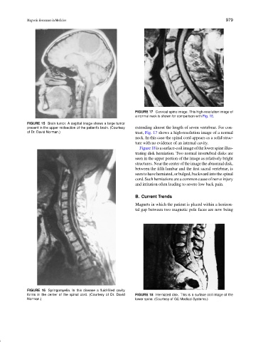
FIGURE 17 Cervical spine image. This high-resolution image of
a normal neck is shown for comparison with Fig. 16.
FIGURE 15 Brain tumor. A sagittal image shows a large tumor
present in the upper midsection of the patient’s brain. (Courtesy extending almost the length of seven vertebrae. For con-
of Dr. David Norman.) trast, Fig. 17 shows a high-resolution image of a normal
neck. In this case the spinal cord appears as a solid struc-
ture with no evidence of an internal cavity.
Figure18isasurface-coilimageofthelowerspineillus-
trating disk herniation. Two normal invertebral disks are
seen in the upper portion of the image as relatively bright
structures. Near the center of the image the abnormal disk,
between the fifth lumbar and the first sacral vertebrae, is
seen to have herniated, or bulged, backward into the spinal
cord. Such herniations are a common cause of nerve injury
and irritation often leading to severe low back pain.
B. Current Trends
Magnets in which the patient is placed within a horizon-
tal gap between two magnetic pole faces are now being
FIGURE 16 Syringomyelia. In this disease a fluid-filled cavity
forms in the center of the spinal cord. (Courtesy of Dr. David FIGURE 18 Herniated disk. This is a surface coil image of the
Norman.) lower spine. (Courtesy of GE Medical Systems.)