
Page 51 - Engineered Interfaces in Fiber Reinforced Composites
P. 51
34 Engineered interfaces in fiber reinforced composites
the specimen, which is influenced by the molecular structure. Therefore, the light
scattering technique provides information about molecular structure and orienta-
tion. Typically, spherulite structures in crystalline polymers are characterized by
complementary SALS and polarized light microscopy, where the scattering angle in
the SALS pattern is used to determine the size of the spherulite. In a similar
approach, SAXS can be used to characterize the structure and dimensions of rigid
fillers or fibers in a thin polymer (Young et al., 1985).
2.3.1 I, Measurement of contact angle
2.3.11.1. Contact angle on aBat surface
Measurements of the contact angle are extremely useful for determining the
wettability of a solid surface by a liquid. Various techniques for measuring the
contact angle have been reviewed by Neumann and Good (1979) and Adamson
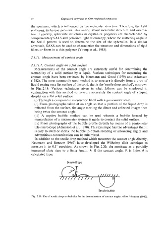
(1982). The most commonly used method is to measure it directly from a drop of
liquid resting on a flat surface of the solid, that is the 'sessile drop method', as shown
in Fig. 2.19. Various techniques given in what follows can be employed in
conjunction with this method to measure accurately the contact angle of a liquid
droplet on a flat solid surface:
(i) Through a comparator microscope filled with a goniometer scale.
(ii) From photographs taken at an angle so that a portion of the liquid drop is
reflected from the surface, the angle meeting the direct and reflected images then
being twice the contact angle.
(iii) A captive bubble method can be used wherein a bubble formed by
manipulation of a micrometer syringe is made to contact the solid surface.
(iv) From photographs of the bubble profile directly by means of a goniometer
tele-microscope (Adamson et al., 1970). This technique has the advantages that it
is easy to swell or shrink the bubble to obtain receding or advancing angles and
adventitious contamination can be minimized.
In addition to the sessile drop method which measures the contact angle directly,
Neumann and Renzow (1 969) have developed the Wilhelmy slide technique to
measure it to 0.1" precision. As shown in Fig. 2.20, the meniscus at a partially
immersed plate rises to a finite length, h, if the contact angle, 8, is finite. 6 is
calculated from
Sessile Drops
Sessite bubble
Fig. 2.19. Use of sessile drops or bubbles for the determination of contact angles. After Adamson (1982).