
Page 354 - Instrumentation Reference Book 3E
P. 354
Mass spectrometers 337
separated by a 10 percent valley, the resolution of dard so that the unknown components are readily
the instrument is 100.00010.005, Le., 20 000. identified and the concentration measured. By
Instrumerits with a resolution of greater than suitable modifications an instrument can be made
150,000 are readily available. The sensitivity. on to provide an energy analysis of electrons released
the other hand, is a measure of the smallest from the surface of a sample by X-radiation, or
detectable quantity of the substance being identi- ultraviolet light.
fied. An example of the extreme sensitivity of
modern instruments is that at a resolution of 16.7.1 Principle of the classical instrument
1000, 3ngk of a compound, relative molecular
mass 300, will give a spectrum with a signal-to- There are many different types of mass spectro-
noise ratio of 10: 1 for a peak having an intensity meters; but the ones described here are the most
of 5 percent of the base peak when a mass range commonly used.
of 1O:l is scanned in 3 s. In all types the pressure is reduced to about
The mass spectrometer has a very wide range 105N/m2 in order to reduce collisions between
of use in process monitoring and laboratory particles in the system. The spectrometer consists
research. It is used in refineries for trace element of an inlet system by which the sample is intro-
survey, analysis of lubricating oils, and identifying duced into the region in which ions of the sample
and quantifying the substances in mixtures of are produced. The separation of ions according to
organic compounds. Its use in detecting and mea- their mass-to-charge ratio may be achieved by
suring the concentration of pollutants in air, magnetic or electric fields or by a combination
water, and solids is rapidly increasing, also its of both. The differences between the various types
use in biochemical analysis in medicine and other of mass spectrometer lie in the manner in which
fields, particularly the analysis of drugs in biolo- the separation is achieved. In the instrument
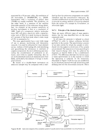
gical extracts. illustrated in Figure 16.20 the ions are accelerated
By means of a double-beam instrument an by an electrical potential through accelerating and
unknown sample may be compared with a stan- defining slits into the electrostatic analyzer, where
to display integrated
charge in peak
Photoplate magazine
analyzer system
Figure 16.28) Schematic diagram of the complete system of a spark source mass spectrometer Courtesy Kratos Ltd