
Page 524 - Instrumentation Reference Book 3E
P. 524
Wavelength and color 507
source whose SPD is known. In the single-beam Both single-beam and double-beam methods
method light from the source is passed through a require the unknown source to remain constant
monochromator to a detector whose output is in intensity whilst its spectrum is scanned-which
recorded at each wavelength of the desired range. can take up to 3 minutes or so. Usually this is not
This is repeated with the source whose SPD is difficult to arrange but if a source is inherently
known, and the ratio of the readings of the two unstable (for example, a carbon arc lamp) the
at each wavelength is then used to determine the whole light output, or output at a single wave-
unknown SPD in relative terms. If the SPD of the length, can be monitored to provide a reference
reference source is known in absolute terms then (Tarrant, 1967).
the SPD of the unknown can be determined in Spectroradiometry is not without its pitfalls,
absolute terms. and the worker intending to embark in the field
In the double-beam method light from the two should consult suitable texts (Forsythe 1941;
sources is passed alternately through a Commission Internationale de I’Eclairage 1984).
monochromator to a detector, enabling the ratio Particular problems arise (1) where line and
of the source outputs to be determined wave- continuous spectra are present together and (2)
length by wavelength. This method is sometimes where the source concerned is tine modulated
offered as an “alternative mode” of using double- (for example, fluorescent lamps, cathode ray
beam spectrophotometers. The experience of the tubes, etc.). When line and continuous spectra
author is that with modern techniques the single- are present together they are transmitted throuzh
beam technique is simpler, more flexible, more the monochromator in differing proportions, and
accurate and as rapid as the double-beam one. this must be taken into account or compensated
It is not usually worthwhile to try to modify a for (Henderson 1970: Moore 1984). The modu-
spectrophotometer to act as a spectroradiometer lation problem can be dealt with by giving the
if good accuracy is sought for. detector a long time constant (which implies a
A recent variation of the single-beam technique slow scan speed) or by the use of phase-locked
is found in the optical multichannel analyzer. detectors (Brown and Tarrant I98 1) (see Section
Here light from the source is dispersed and made 21.4.3).
to fall not on a slit but on a multiple-array detec- Few firms offer spectroradiometers as stock
tor; each detector bit is read separately with the lines because they nearly always have to be cus-
aid of a microprocessor. This technique is not as tom built for particular applications. One recent
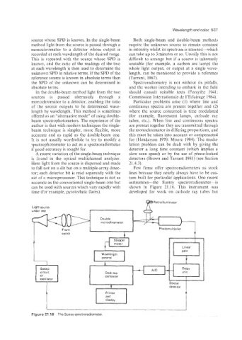
accurate as the conventional single-beam one but instrument-the Surrey spectroradiometer-is
can be used with sources which vary rapidly with shown in Figure 21.16. This instrument was
time (for example, pyrotechnic flares). developed for work on cathode ray tubes but
T
Retroilluminator
Light source
under test
Double
monochromator
Front Photomultiplier
optics
control
I J
Figure 21.1 6 The Surreyspectroradiometer.