Page 267 -
P. 267
4-8                                                              MEMS: Design and Fabrication


             superconducting materials can be made in bulk, compact storage rings will become extremely attractive.
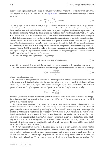
             The angular opening of the radiation cone in Figure 4.4 is determined by the electron energy, E, and is
             given by:
                                                    mc 2     0.5
                                               θ                   (mrad)                               (4.1)
                                                     E     E(GeV)

             The X-ray light bundle with the cone opening, θ, describes a horizontal line on an intersecting substrate
             as the X-ray bundle is tangent to the circular electron path. In the vertical direction, the intensity of the
             beam exhibits a Gaussian distribution, and the vertical exposed height on the intersecting substrate can
             be calculated knowing θ and R, the distance from the radiation point P to the substrate. With E   1GeV,
             θ   1 mrad, and R   10 m, the exposed area in the vertical direction measures about 0.5cm. To expose
             a substrate homogeneously over a wider vertical range, the sample is moved vertically through the irra-
             diation band with a precision scanner, for example, at a speed of 10 mm/s over a 100mm scanning dis-
             tance. Usually, the substrate is stepped up and down repeatedly until the desired X-ray dose is obtained.
             It is interesting to note that an SOR setup affords continuous lithography, a prospect that may make dis-
             posable ICs and MEMS a possibility. Rolls of dry X-ray photoresist or X-ray photoresist-covered foils
             could pass through the exposure beam, resulting in a continuous lithography process — that is, a “beyond
             batch” type of approach (see inset in Figure 4.4).
               The electron energy E in Equation (4.1) is given by:


                                           E(GeV)   0.29979B(Tesla)ρ(meters)                            (4.2)

             where B is the magnetic field and ρ is the radius of the circular path of the electrons in the synchrotron.
               The total radiated power can be calculated from the energy loss of the electrons per turn and is given by:
                                                                   4
                                                             88.47E i
                                                   P(kW)                                                (4.3)
                                                                ρ
             where i is the beam current.
               The emission of the synchrotron electrons is a broad spectrum without characteristic peaks or line
             enhancements, and  its  distribution  extends  from  the  microwave  region  through  the  infrared, visible,
             ultraviolet, and into the X-ray region. The critical wavelength, λ , is defined so that the total radiated
                                                                         c
             power at lower wavelengths equals the radiated power at higher wavelengths, and is given by:
                                                            5.59ρ(m)
                                                   λ (Å)                                                (4.4)
                                                              3
                                                    c       E (GeV)
             Equation (4.3) shows that the total radiated power increases with the fourth power of the electron energy.
             From Equation (4.4), we appreciate that the spectrum shifts toward shorter wavelengths with the third
             power of the electron energy.
               The dose variation absorbed in the top vs. the bottom of an X-ray resist should be kept small so that
             the top layer does not deteriorate before the bottom layers are sufficiently exposed. Since the depth of
             penetration increases with decreasing wavelength, synchrotron radiation of very short wavelengths is
             needed to pattern thick resist layers. To obtain good aspect ratios in LIGA structures, the critical wave-
             length ideally should be 2Å. Bley et al. (1992) at KfK designed a new synchrotron optimized for LIGA.
             They proposed a magnetic flux density, B, of 1.6285 T; a nominal energy, E, of 2.3923GeV; and a bend-
             ing radius, ρ, of 4.9 m. With those parameters, Equation (4.4) results in the desired λ of 2Å and an open-
                                                                                         c
             ing angle of radiation, based on Equation (4.1), of 0.2 mrad (in practice, this angle will be closer to 0.3
             mrad due to electron beam emittance).
               The X-rays traveling from the ring to the sample site are held in a high vacuum. The sample itself is
             kept either in air or in a helium (He) atmosphere. The inert atmosphere prevents corrosion of the expo-
             sure chamber, mask, and sample by reactive oxygen species, and removal of heat is much faster than in
             air (the heat conductivity of He is high compared to air). In He, the X-ray intensity loss is also 500 times
             less than in air. A beryllium (Be) window separates the high vacuum from the inert atmosphere. For


             © 2006 by Taylor & Francis Group, LLC

