
Page 265 -
P. 265
4-6 MEMS: Design and Fabrication
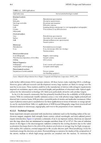
TABLE 4.2 SOR Applications
Application area Instruments/technologies needed
Structural analysis
Atoms Photoelectron spectrometers
Molecules Absorption spectrometers
Very large molecules Fluorescent spectrometers
Proteins Diffraction cameras
Cells Scanning electron microscope (to view topographical radiographs)
Crystals Time resolved X-ray diffractometers
Polycrystals
Chemical analysis
Trace Photoelectron spectrometers
Surface (Secondary ion) mass spectrometer
Bulk Absorption/fluorescence spectrometers
Vacuum systems
Microscopy
Photoelectron Photoemission microscopes
X-ray X-ray microscopes SEM (for viewing)
Vacuum systems
Micro/nanofabrication
X-ray lithography Steppers, mask making
Photochemical deposition of thin films Vacuum systems
Etching LIGA process
Medical diagnostics
Radiography X-ray cameras and equipment
Angiography and tomography Computer aided display
Photochemical reactions
Preparation of novel materials Vacuum systems
Gas-handling equipment
Source: Adapted with permission from Nippon Telegraph and Telephone Corporation (NNT), 1991.
walls further differentiates LIGA exposure stations. All these factors make exploring LIGA a challenge.
However, given sufficient research and development money, large markets are likely to emerge over the
next five to ten years. These markets could be in the manufacture of devices with stringent requirements
imposed on resolution, aspect ratio, structural height, and parallelism of structural walls. Optical appli-
cations for the information technology (IT) field seem particularly attractive early product targets.
So far, it is the research community that has primarily benefited from the availability of SOR photon
sources. With its continuously tunable radiation across a very wide photon range, highly polarized and
directed into a narrow beam, SOR provides a powerful probe of atomic and molecular resonances. Other
types of photon sources prove unsatisfactory for these applications in terms of intensity or energy spread.
As can be concluded from Table 4.2, applications of SOR beyond lithography range from structural and
chemical analysis to microscopy, angiography, and even to the preparation of new materials.
4.2.2.2 Technical Aspects
Some important concepts associated with synchrotron radiation (such as the bending radius of the syn-
chrotron magnet, magnetic field strength, beam current, critical wavelength, and total radiated power)
require introduction. Figure 4.4 presents a schematic of an X-ray exposure station. Electrons are injected
9
6
into the ring, where they are maintained at energies anywhere from 10 to 10 eV. The cone of radiation
shown in this figure is the electromagnetic radiation emitted by the circling electrons due to the radial
acceleration that keeps them in the orbit of the electron synchrotron or storage ring. For high-energy par-
ticle studies, this radiation, emitted tangential to the circular electron path (Bremsstrahlung), limits the
maximum energy the electrons can attain. Bremsstrahlung is a nuisance for studies of the composition of
the atomic nucleus in which high-energy particles are smashed into the nucleus. To minimize the
© 2006 by Taylor & Francis Group, LLC