
Page 181 - Science at the nanoscale
P. 181
RPS: PSP0007 - Science-at-Nanoscale
10:16
June 5, 2009
8.2. Electron Microscopy
(AE). The AE has a characteristic energy, unique to each element
from which it was emitted. AE has relatively low energy and are
only emitted from the surface of the specimen, typically from a
depth of <3 nm, thereby yielding surface sensitive compositional
information. Hence, both EDX and Auger electron spectroscopy
are used for elemental analysis or chemical characterisation of a
sample.
Light is also emitted when a sample is being bombarded with
electrons. Many substances give out light when bombarded with
electrons, just like a TV monitor. This effect can be exploited for
imaging. The light emitted can be in the ultra-violet, visible or
infrared range and this phenomenon is referred to as Catholumi-
nescence (CL).
The SEM generates an image of the sample by scanning the elec-
trons over the sample surface while a SE detector placed near
the sample collects the signal generated. Modern SEMs have
incorporated many attractive technical features so that imaging
with a SEM has become very user friendly. The images are gen-
erated almost real time and high quality images can be stored
directly in digital format. A simple turn of a knob allows us to
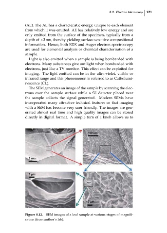
Figure 8.12. SEM images of a leaf sample at various stages of magnifi- 171 ch08
cation (from author’s lab).