
Page 183 - Science at the nanoscale
P. 183
10:16
June 5, 2009
8.2. Electron Microscopy
High Resolution TEM (HRTEM) is now routinely used to achieve
atomic resolution of a sample. However, TEM has its limitations.
Lengthy sample preparation is usually required to make the sam-
ple thin enough. Since the beam is traveling through the sample,
the sample bulk and not the surface is being imaged.
How does a TEM work?
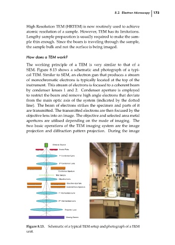
The working principle of a TEM is very similar to that of a
SEM. Figure 8.13 shows a schematic and photograph of a typi-
cal TEM. Similar to SEM, an electron gun that produces a stream
of monochromatic electrons is typically located at the top of the
instrument. This stream of electrons is focused to a coherent beam
by condenser lenses 1 and 2. Condenser aperture is employed
to restrict the beam and remove high angle electrons that deviate
from the main optic axis of the system (indicated by the dotted
line). The beam of electrons strikes the specimen and parts of it
are transmitted. The transmitted electrons are then focused by the
objective lens into an image. The objective and selected area metal
apertures are utilised depending on the mode of imaging. The
two basic operations of the TEM imaging system are the image
projection and diffraction pattern projection. During the image
Electron Source
Anode Plate
1 st Condenser Lens
2 nd Condenser Lens
Condenser Aperture RPS: PSP0007 - Science-at-Nanoscale 173 ch08
Thin Sample
Objective Lens
Objective Aperture
Selected Area Aperture
1 st Intermediate Lens
2 nd Intermediate Lens
Projector Lens
Viewing Screen
------------------------------------------------------------------------------------
Figure 8.13. Schematic of a typical TEM setup and photograph of a TEM
unit.