
Page 240 - Vibrational Spectroscopic Imaging for Biomedical Applications
P. 240
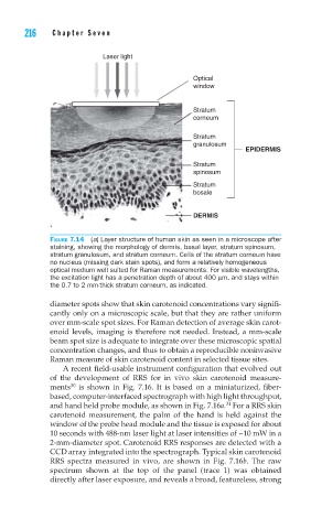
216    Cha pte r  Se v e n


                      Laser light

                                            Optical
                                            window


                                            Stratum
                                            corneum

                                            Stratum
                                            granulosum
                                                         EPIDERMIS

                                            Stratum
                                            spinosum
                                            Stratum
                                            bosale


                                            DERMIS

        FIGURE 7.14 (a) Layer structure of human skin as seen in a microscope after
        staining, showing the morphology of dermis, basal layer, stratum spinosum,
        stratum granulosum, and stratum corneum. Cells of the stratum corneum have
        no nucleus (missing dark stain spots), and form a relatively homogeneous
        optical medium well suited for Raman measurements. For visible wavelengths,
        the excitation light has a penetration depth of about 400 μm, and stays within
        the 0.7 to 2 mm-thick stratum corneum, as indicated.

        diameter spots show that skin carotenoid concentrations vary signifi-
        cantly only on a microscopic scale, but that they are rather uniform
        over mm-scale spot sizes. For Raman detection of average skin carot-
        enoid levels, imaging is therefore not needed. Instead, a mm-scale
        beam spot size is adequate to integrate over these microscopic spatial
        concentration changes, and thus to obtain a reproducible noninvasive
        Raman measure of skin carotenoid content in selected tissue sites.
            A recent field-usable instrument configuration that evolved out
        of the development of RRS for in vivo skin carotenoid measure-
              30
        ments  is shown in Fig. 7.16. It is based on a miniaturized, fiber-
        based, computer-interfaced spectrograph with high light throughput,
                                                    31
        and hand held probe module, as shown in Fig. 7.16a.  For a RRS skin
        carotenoid measurement, the palm of the hand is held against the
        window of the probe head module and the tissue is exposed for about
        10 seconds with 488-nm laser light at laser intensities of ~10 mW in a
        2-mm-diameter spot. Carotenoid RRS responses are detected with a
        CCD array integrated into the spectrograph. Typical skin carotenoid
        RRS spectra measured in vivo, are shown in Fig. 7.16b. The raw
        spectrum shown at the top of the panel (trace 1) was obtained
        directly after laser exposure, and reveals a broad, featureless, strong